• 文献检索
  • 文档翻译
  • 深度研究
  • 学术资讯
  • Suppr Zotero 插件Zotero 插件
  • 邀请有礼
  • 套餐&价格
  • 历史记录
应用&插件
Suppr Zotero 插件Zotero 插件浏览器插件Mac 客户端Windows 客户端微信小程序
定价
高级版会员购买积分包购买API积分包
服务
文献检索文档翻译深度研究API 文档MCP 服务
关于我们
关于 Suppr公司介绍联系我们用户协议隐私条款
关注我们

Suppr 超能文献

核心技术专利:CN118964589B侵权必究
粤ICP备2023148730 号-1Suppr @ 2026

文献检索

告别复杂PubMed语法,用中文像聊天一样搜索,搜遍4000万医学文献。AI智能推荐,让科研检索更轻松。

立即免费搜索

文件翻译

保留排版,准确专业,支持PDF/Word/PPT等文件格式,支持 12+语言互译。

免费翻译文档

深度研究

AI帮你快速写综述,25分钟生成高质量综述,智能提取关键信息,辅助科研写作。

立即免费体验

应用扫频源和谱域光学相干断层扫描测量肥厚脉络膜疾病脉络膜厚度的比较。

Comparison of choroidal thickness measurements using swept source and spectral domain optical coherence tomography in pachychoroid diseases.

机构信息

Department of Ophthalmology, Konyang University College of Medicine, Daejeon, Republic of Korea.

Department of Ophthalmology, Chungnam National University College of Medicine, Daejeon, Republic of Korea.

出版信息

PLoS One. 2020 Feb 26;15(2):e0229134. doi: 10.1371/journal.pone.0229134. eCollection 2020.

DOI:10.1371/journal.pone.0229134
PMID:32101541
原文链接:https://pmc.ncbi.nlm.nih.gov/articles/PMC7043756/
Abstract

PURPOSE

To determine the comparability of choroidal thickness (ChT) measurements using swept source (SS) and spectral domain (SD) optical coherence tomography (OCT) devices in patients with pachychoroid diseases.

METHODS

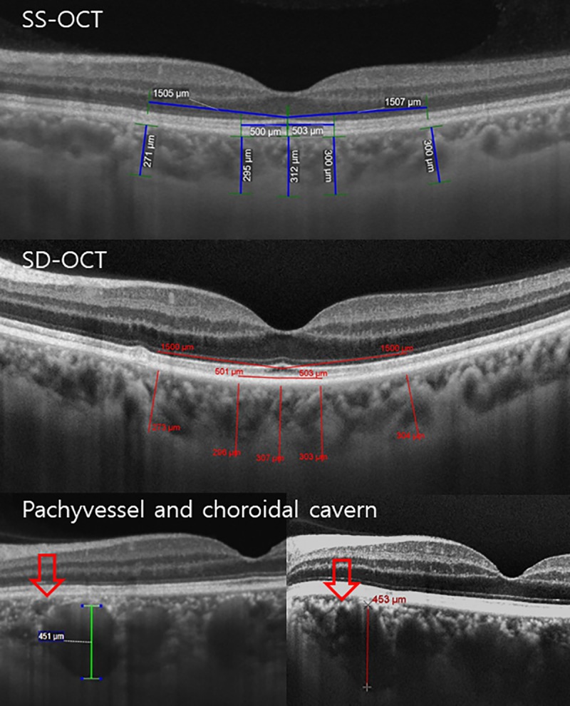
Patients with pachychoroid diseases were recruited. OCT scans were performed sequentially with a Cirrus HD OCT 5000 and Plex Elite 9000. Images were analyzed by two independent observers. Each image was independently measured twice by each observer to determine the intraobserver repeatability.

RESULTS

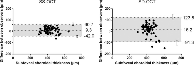
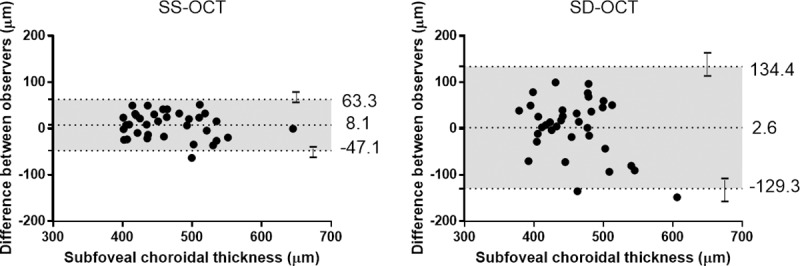
A total of 55 eyes were included. The average ChT of the subfoveal area using SS-OCT and SD-OCT was 430.5 ± 68.1 and 428.5 ± 57.9 μm, respectively, which did not show a significant result as the main effect in the repeated-measure analysis of variance (P = 0.067). Using SS-OCT, the intraobserver intraclass correlation coefficient (ICC) of both observers was > 0.950 at every measured point, and the interobserver coefficient of repeatability (CR) of the subfoveal area was 45.1 μm (95% confidence interval (CI), 40.8-49.4). Using SD-OCT, the intraobserver ICC of both observers was > 0.800, and the interobserver CR of the subfoveal area was 71.2 μm (95% CI, 64.4-78.0). Additionally, the intraobserver and interobserver CRs showed significantly better repeatability in SS-OCT than SD-OCT in F-test. In patients with ChT ≥ 400 μm, the interobserver CRs of SS-OCT and SD-OCT were 48.4 (95% CI, 42.6-54.2) and 95.2 μm (95% CI, 83.9-106.6), respectively. In patients with a subfoveal active lesion, the interobserver CRs were 44.5 (95% CI, 37.6-51.4) and 100.1 μm (95% CI, 84.6-115.5), respectively.

CONCLUSIONS

Although the ChT measurements were comparable between SS-OCT and SD-OCT devices in pachychoroid diseases, SD-OCT showed low reliability in patients with ChT ≥ 400 μm and subfoveal active lesions. SS-OCT would be therefore more suitable for observation and follow-up of choroidal structures in pachychoroid diseases.

摘要

目的

确定在患有肥厚脉络膜疾病的患者中,使用扫频源(SS)和谱域(SD)光学相干断层扫描(OCT)设备测量脉络膜厚度(ChT)的可比性。

方法

招募患有肥厚脉络膜疾病的患者。使用 Cirrus HD OCT 5000 和 Plex Elite 9000 连续进行 OCT 扫描。由两位独立观察者对图像进行分析。每位观察者独立对每张图像进行两次测量,以确定观察者内的重复性。

结果

共纳入 55 只眼。SS-OCT 和 SD-OCT 测量的中心凹下区域平均 ChT 分别为 430.5±68.1μm 和 428.5±57.9μm,重复测量方差分析的主要效应无显著差异(P=0.067)。使用 SS-OCT,两位观察者的每个测量点的观察者内组内相关系数(ICC)均>0.950,中心凹下区域的观察者间重复性可重复性系数(CR)为 45.1μm(95%置信区间(CI),40.8-49.4)。使用 SD-OCT,两位观察者的观察者内 ICC 均>0.800,中心凹下区域的观察者间 CR 为 71.2μm(95%CI,64.4-78.0)。此外,F 检验显示,在 SS-OCT 中,观察者内和观察者间的 CR 比 SD-OCT 具有更好的重复性。在 ChT≥400μm 的患者中,SS-OCT 和 SD-OCT 的观察者间 CR 分别为 48.4μm(95%CI,42.6-54.2)和 95.2μm(95%CI,83.9-106.6)。在中心凹下有活动性病变的患者中,观察者间 CR 分别为 44.5μm(95%CI,37.6-51.4)和 100.1μm(95%CI,84.6-115.5)。

结论

尽管在肥厚脉络膜疾病中 SS-OCT 和 SD-OCT 设备的 ChT 测量值具有可比性,但在 ChT≥400μm 和中心凹下有活动性病变的患者中,SD-OCT 的可靠性较低。因此,SS-OCT 更适合于肥厚脉络膜疾病脉络膜结构的观察和随访。

https://cdn.ncbi.nlm.nih.gov/pmc/blobs/834f/7043756/c8e578df4f4a/pone.0229134.g004.jpg
https://cdn.ncbi.nlm.nih.gov/pmc/blobs/834f/7043756/d6d30e97ae93/pone.0229134.g001.jpg
https://cdn.ncbi.nlm.nih.gov/pmc/blobs/834f/7043756/fac48b6a777c/pone.0229134.g002.jpg
https://cdn.ncbi.nlm.nih.gov/pmc/blobs/834f/7043756/730a6e35ba3b/pone.0229134.g003.jpg
https://cdn.ncbi.nlm.nih.gov/pmc/blobs/834f/7043756/c8e578df4f4a/pone.0229134.g004.jpg

相似文献

1
Comparison of choroidal thickness measurements using swept source and spectral domain optical coherence tomography in pachychoroid diseases.应用扫频源和谱域光学相干断层扫描测量肥厚脉络膜疾病脉络膜厚度的比较。
PLoS One. 2020 Feb 26;15(2):e0229134. doi: 10.1371/journal.pone.0229134. eCollection 2020.
2
Comparison of choroidal thickness measurements between spectral domain optical coherence tomography and swept source optical coherence tomography in children.比较儿童脉络膜厚度测量在光谱域光学相干断层扫描和扫频源光学相干断层扫描之间的差异。
Sci Rep. 2021 Jul 2;11(1):13749. doi: 10.1038/s41598-021-92980-9.
3
Comparison of choroidal thicknesses using swept source and spectral domain optical coherence tomography in diseased and normal eyes.使用扫频源光学相干断层扫描和光谱域光学相干断层扫描对患病眼和正常眼的脉络膜厚度进行比较。
Br J Ophthalmol. 2015 Mar;99(3):354-8. doi: 10.1136/bjophthalmol-2014-305331. Epub 2014 Oct 1.
4
Direct comparison of spectral-domain and swept-source OCT in the measurement of choroidal thickness in normal eyes.直接比较谱域和扫频源光学相干断层扫描在正常眼脉络膜厚度测量中的应用。
Br J Ophthalmol. 2014 Mar;98(3):334-8. doi: 10.1136/bjophthalmol-2013-303904. Epub 2013 Nov 28.
5
Comparisons of choroidal thickness of normal eyes obtained by two different spectral-domain OCT instruments and one swept-source OCT instrument.两种不同的谱域 OCT 仪器和一种扫频源 OCT 仪器测量正常眼脉络膜厚度的比较。
Invest Ophthalmol Vis Sci. 2013 Nov 19;54(12):7630-6. doi: 10.1167/iovs.13-13135.
6
Comparison of choroidal thickness using swept-source and spectral-domain optical coherence tomography in normal Indian eyes.在正常印度人眼中使用扫频源光学相干断层扫描和谱域光学相干断层扫描比较脉络膜厚度
Oman J Ophthalmol. 2018 Jan-Apr;11(1):38-41. doi: 10.4103/ojo.OJO_27_2017.
7
A comparison of choroidal thicknesses between pachychoroid and normochoroid eyes acquired from wide-field swept-source OCT.宽视野扫频源光学相干断层扫描获得的厚脉络膜与正常脉络膜眼中脉络膜厚度的比较。
Acta Ophthalmol. 2021 Feb;99(1):e117-e123. doi: 10.1111/aos.14522. Epub 2020 Jun 22.
8
REAL-TIME FULL-DEPTH VISUALIZATION OF POSTERIOR OCULAR STRUCTURES: Comparison Between Full-Depth Imaging Spectral Domain Optical Coherence Tomography and Swept-Source Optical Coherence Tomography.眼后段结构的实时全深度可视化:全深度成像光谱域光学相干断层扫描与扫频源光学相干断层扫描的比较
Retina. 2016 Jun;36(6):1153-61. doi: 10.1097/IAE.0000000000000842.
9
Repeatability of manual subfoveal choroidal thickness measurements in healthy subjects using the technique of enhanced depth imaging optical coherence tomography.应用增强深度成像光学相干断层扫描技术测量健康受试者的手动黄斑中心凹下脉络膜厚度的重复性。
Invest Ophthalmol Vis Sci. 2011 Apr 8;52(5):2267-71. doi: 10.1167/iovs.10-6024. Print 2011 Apr.
10
Comparison of peripapillary and subfoveal choroidal thickness in normal versus primary open-angle glaucoma (POAG) subjects using spectral domain optical coherence tomography (SD-OCT) and swept source optical coherence tomography (SS-OCT).使用频域光学相干断层扫描(SD-OCT)和扫频光学相干断层扫描(SS-OCT)比较正常人和原发性开角型青光眼(POAG)患者视乳头周围和黄斑中心凹下脉络膜厚度。
BMJ Open Ophthalmol. 2019 Jul 11;4(1):e000258. doi: 10.1136/bmjophth-2018-000258. eCollection 2019.

引用本文的文献

1
Repeatability, reproducibility, and agreement for three optical coherence tomography instruments.三种光学相干断层扫描仪器的可重复性、再现性及一致性
BMC Ophthalmol. 2025 Jul 28;25(1):427. doi: 10.1186/s12886-025-04257-8.
2
Imaging in Pachychoroid Disease.肥厚性脉络膜疾病的影像学检查
Turk J Ophthalmol. 2025 Feb 27;55(1):36-48. doi: 10.4274/tjo.galenos.2024.40388. Epub 2025 Feb 5.
3
Potential Drug Targets for Diabetic Retinopathy Identified Through Mendelian Randomization Analysis.通过孟德尔随机化分析鉴定出的糖尿病性视网膜病变的潜在药物靶点。

本文引用的文献

1
Repeatability of wide-field choroidal thickness measurements using enhanced-depth imaging optical coherence tomography.使用增强深度成像光学相干断层扫描测量宽视野脉络膜厚度的可重复性
Clin Exp Optom. 2019 May;102(3):327-334. doi: 10.1111/cxo.12893. Epub 2019 Mar 18.
2
Measurable Range of Subfoveal Choroidal Thickness With Conventional Spectral Domain Optical Coherence Tomography.传统光谱域光学相干断层扫描测量黄斑中心凹下脉络膜厚度的可测量范围
Transl Vis Sci Technol. 2018 Oct 1;7(5):16. doi: 10.1167/tvst.7.5.16. eCollection 2018 Sep.
3
Subfoveal choroidal thickness as a prognostic factor in exudative age-related macular degeneration.
Transl Vis Sci Technol. 2024 Nov 4;13(11):17. doi: 10.1167/tvst.13.11.17.
4
Segmentation of choroidal area in optical coherence tomography images using a transfer learning-based conventional neural network: a focus on diabetic retinopathy and a literature review.基于迁移学习的传统神经网络在光学相干断层扫描图像中脉络膜区域的分割:以糖尿病视网膜病变为重点并进行文献回顾。
BMC Med Imaging. 2024 Oct 18;24(1):281. doi: 10.1186/s12880-024-01459-2.
5
Comparison of manual and artificial intelligence-automated choroidal thickness segmentation of optical coherence tomography imaging in myopic adults.近视成年人中光学相干断层扫描成像的手动与人工智能自动脉络膜厚度分割比较
Eye Vis (Lond). 2024 Jun 3;11(1):21. doi: 10.1186/s40662-024-00385-2.
6
Impact of Hemodialysis on Subfoveal Choroidal Thickness Measured by Optical Coherence Tomography: A Systematic Review and a Pooled Analysis of Self-Controlled Case Series.血液透析对光学相干断层扫描测量的黄斑中心凹下脉络膜厚度的影响:一项系统评价及自身对照病例系列的汇总分析
Ophthalmol Ther. 2023 Oct;12(5):2265-2280. doi: 10.1007/s40123-023-00761-6. Epub 2023 Jul 13.
7
The Role of Diabetic Choroidopathy in the Pathogenesis and Progression of Diabetic Retinopathy.糖尿病性脉络膜病变在糖尿病性视网膜病变发病机制和进展中的作用。
Int J Mol Sci. 2023 Jun 15;24(12):10167. doi: 10.3390/ijms241210167.
8
Focal Choroidal Excavation: Epidemiology, Clinical Characteristics and Multimodal Imaging Findings.局限性脉络膜凹陷:流行病学、临床特征及多模态影像学表现
Diagnostics (Basel). 2023 Feb 7;13(4):602. doi: 10.3390/diagnostics13040602.
9
Choroidal Neovascularization Is Common in Best Vitelliform Macular Dystrophy and Plays a Role in Vitelliform Lesion Evolution.脉络膜新生血管在 Best 型类卵黄样黄斑营养不良中很常见,并且在类卵黄样病变的演变中起作用。
Ophthalmol Retina. 2023 May;7(5):441-449. doi: 10.1016/j.oret.2022.11.014. Epub 2022 Dec 14.
10
Choroidal Vasculature Changes in Age-Related Macular Degeneration: From a Molecular to a Clinical Perspective.年龄相关性黄斑变性的脉络膜血管变化:从分子到临床角度。
Int J Mol Sci. 2022 Oct 9;23(19):12010. doi: 10.3390/ijms231912010.
黄斑部下脉络膜厚度在渗出性老年黄斑部变性的预后因子。
Br J Ophthalmol. 2019 Jul;103(7):918-921. doi: 10.1136/bjophthalmol-2018-312625. Epub 2018 Aug 27.
4
Choroidal thickness maps from spectral domain and swept source optical coherence tomography: algorithmic versus ground truth annotation.光谱域和扫频源光学相干断层扫描的脉络膜厚度图:算法标注与真实标注对比
Br J Ophthalmol. 2016 Oct;100(10):1372-6. doi: 10.1136/bjophthalmol-2015-307985. Epub 2016 Jan 14.
5
Comparison of macular choroidal thicknesses from swept source and spectral domain optical coherence tomography.扫频源光学相干断层扫描与谱域光学相干断层扫描测量黄斑脉络膜厚度的比较
Br J Ophthalmol. 2016 Jul;100(7):995-999. doi: 10.1136/bjophthalmol-2015-307541. Epub 2015 Nov 9.
6
EN FACE IMAGING OF PACHYCHOROID SPECTRUM DISORDERS WITH SWEPT-SOURCE OPTICAL COHERENCE TOMOGRAPHY.使用扫频光学相干断层扫描对厚脉络膜疾病谱进行正面成像
Retina. 2016 Mar;36(3):499-516. doi: 10.1097/IAE.0000000000000742.
7
Pachychoroid diseases of the macula.黄斑部厚脉络膜疾病
Med Hypothesis Discov Innov Ophthalmol. 2014 Winter;3(4):111-5.
8
Exact parametric confidence intervals for Bland-Altman limits of agreement.布兰德-奥特曼一致性界限的精确参数置信区间。
Optom Vis Sci. 2015 Mar;92(3):e71-80. doi: 10.1097/OPX.0000000000000513.
9
Comparison of penetration depth in choroidal imaging using swept source vs spectral domain optical coherence tomography.扫频源光学相干断层扫描与谱域光学相干断层扫描在脉络膜成像中穿透深度的比较。
Eye (Lond). 2015 Mar;29(3):409-15. doi: 10.1038/eye.2014.319. Epub 2015 Jan 16.
10
Enhanced depth imaging-OCT of the choroid: a review of the current literature.脉络膜增强深度成像光学相干断层扫描:当前文献综述
Graefes Arch Clin Exp Ophthalmol. 2014 Dec;252(12):1871-83. doi: 10.1007/s00417-014-2840-y. Epub 2014 Nov 4.