Yasuda Atsushi, Yasuda Takushi, Imamoto Haruhiko, Hiraki Yoko, Momose Kohta, Kato Hiroaki, Iwama Mitsuru, Shiraishi Osamu, Shinkai Masayuki, Imano Motohiro, Kimura Yutaka
Department of Surgery, Kindai University Faculty of Medicine, 377-2 Ohno-higashi, Osaka-Sayama, Osaka, 589-8511, Japan.
Cancer Center, Kindai University Hospital, 377-2 Ohno-higashi, Osaka-Sayama, Osaka, 589-8511, Japan.
Surg Case Rep. 2020 Feb 27;6(1):44. doi: 10.1186/s40792-020-00809-w.
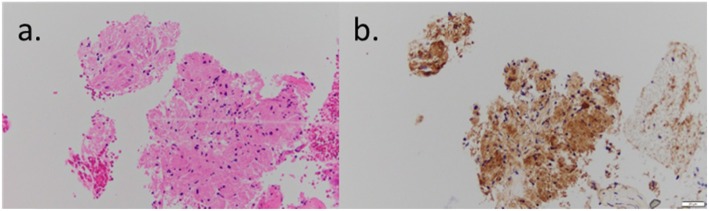
Granular cell tumors (GCT) in the gastrointestinal tract are rare. Herein, we describe a case of a gastric GCT diagnosed preoperatively by endoscopic ultrasound-guided fine needle aspiration biopsy (EUS-FNAB) and successfully resected by single-incision laparoscopic surgery (SILS).
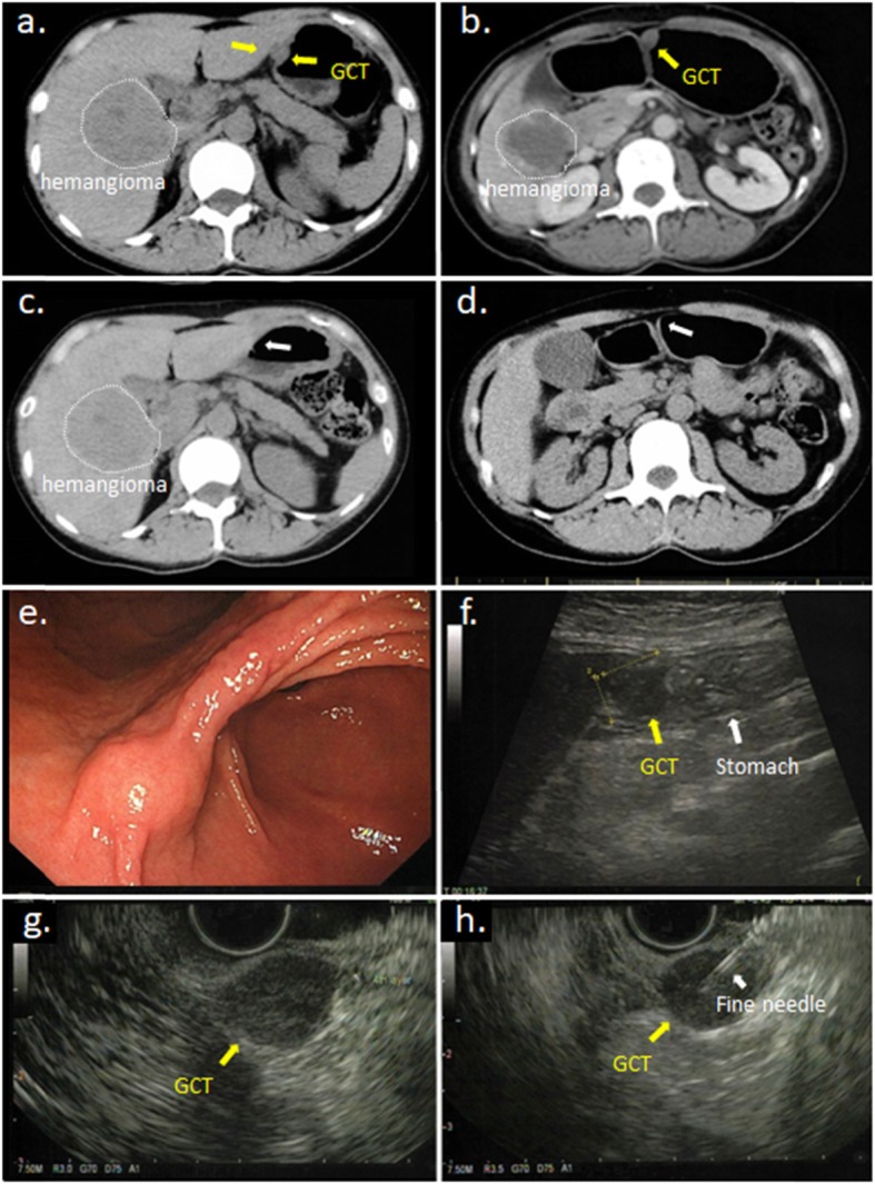
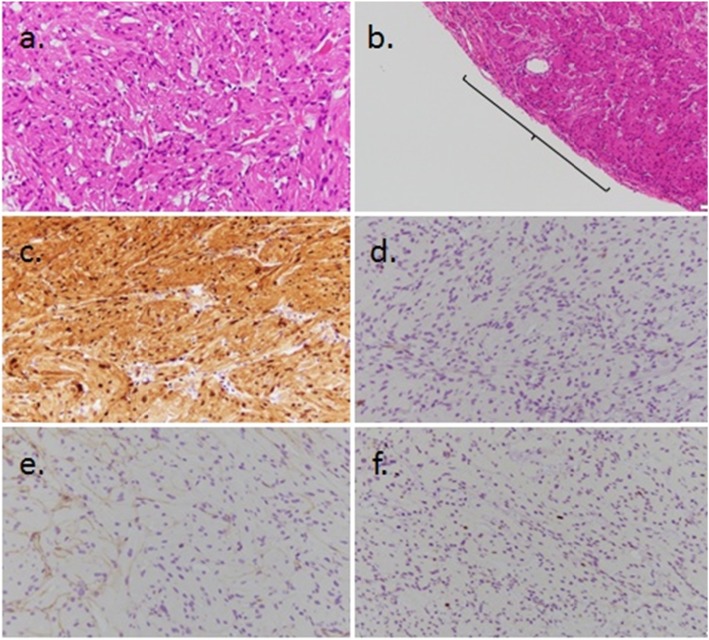
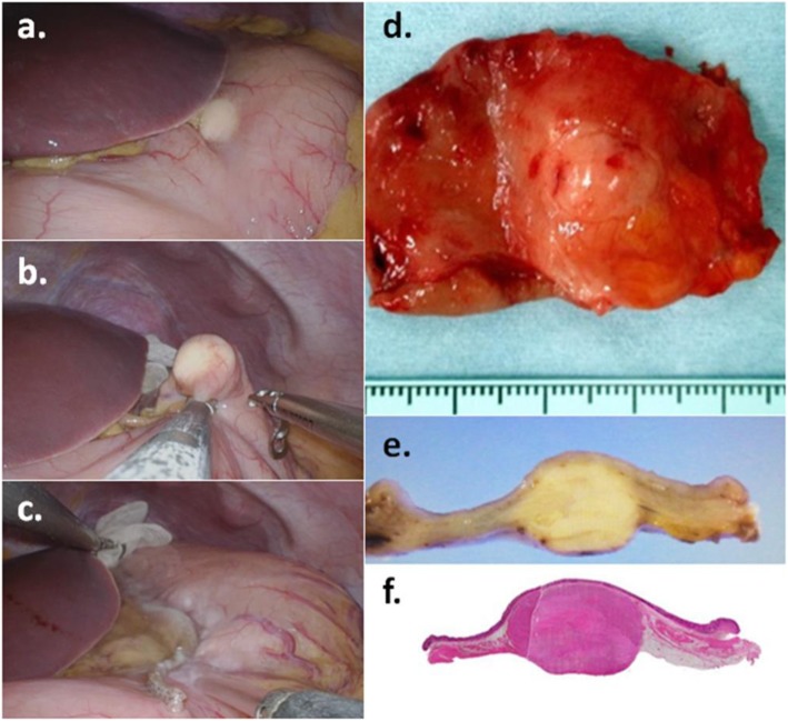
A 46-year-old Japanese woman had a tumor located in the angle of the stomach that was approximately 1.5 cm in diameter. Abdominal computed tomography (CT) revealed a submucosal tumor (SMT), which was finally diagnosed as a gastric GCT using EUS-FNAB. The tumor was not identified by CT 1 year and 4 months before diagnosis; therefore, because there was a possibility that the tumor was malignant, we performed surgical wedge resection using SILS. The patient had an uneventful recovery postoperatively and was discharged without complications 3 days after surgery. The tumor was pathologically diagnosed as a benign GCT that remained within the muscular layer. No recurrence or complications have occurred in the first 16 months since the surgery.
Because gastric GCTs are generally benign and are rarely associated with lymph node metastasis, SILS seems to be a safe and feasible surgical approach for treating GCTs.
胃肠道颗粒细胞瘤(GCT)较为罕见。在此,我们描述一例通过内镜超声引导下细针穿刺活检(EUS-FNAB)术前诊断为胃GCT,并经单孔腹腔镜手术(SILS)成功切除的病例。
一名46岁的日本女性,胃角处有一个直径约1.5厘米的肿瘤。腹部计算机断层扫描(CT)显示为黏膜下肿瘤(SMT),最终通过EUS-FNAB诊断为胃GCT。在诊断前1年4个月的CT检查中未发现该肿瘤;因此,鉴于该肿瘤有恶性的可能性,我们采用SILS进行了手术楔形切除。患者术后恢复顺利,术后3天无并发症出院。肿瘤经病理诊断为良性GCT,局限于肌层内。自手术以来的前16个月未发生复发或并发症。
由于胃GCT通常为良性,很少发生淋巴结转移,SILS似乎是治疗GCT的一种安全可行的手术方法。