Tward Daniel, Brown Timothy, Kageyama Yusuke, Patel Jaymin, Hou Zhipeng, Mori Susumu, Albert Marilyn, Troncoso Juan, Miller Michael
Department of Biomedical Engineering, Johns Hopkins University, Baltimore, MD, United States.
Center for Imaging Science, Johns Hopkins University, Baltimore, MD, United States.
Front Neurosci. 2020 Feb 11;14:52. doi: 10.3389/fnins.2020.00052. eCollection 2020.
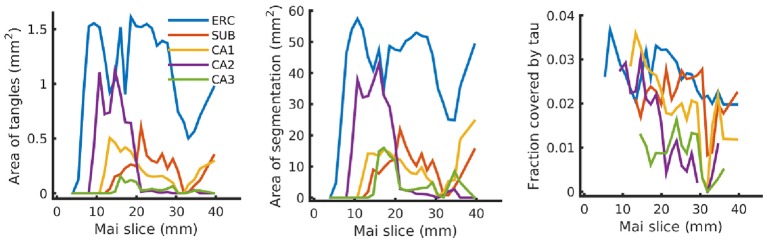
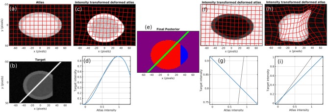
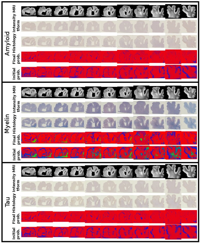
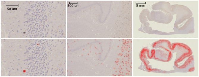
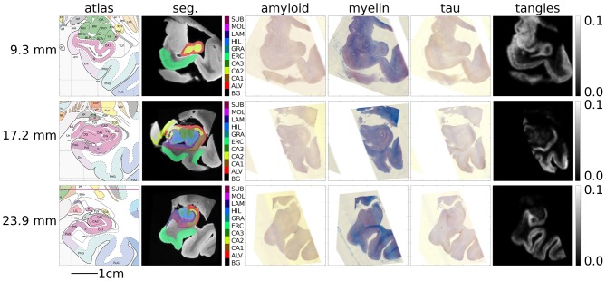
This paper examines the problem of diffeomorphic image registration in the presence of differing image intensity profiles and sparsely sampled, missing, or damaged tissue. Our motivation comes from the problem of aligning 3D brain MRI with 100-micron isotropic resolution to histology sections at 1 × 1 × 1,000-micron resolution with multiple varying stains. We pose registration as a penalized Bayesian estimation, exploiting statistical models of image formation where the target images are modeled as sparse and noisy observations of the atlas. In this injective setting, there is no assumption of symmetry between atlas and target. Cross-modality image matching is achieved by jointly estimating polynomial transformations of the atlas intensity. Missing data is accommodated via a multiple atlas selection procedure where several atlas images may be of homogeneous intensity and correspond to "background" or "artifact." The two concepts are combined within an Expectation-Maximization algorithm, where atlas selection posteriors and deformation parameters are updated iteratively and polynomial coefficients are computed in closed form. We validate our method with simulated images, examples from neuropathology, and a standard benchmarking dataset. Finally, we apply it to reconstructing digital pathology and MRI in standard atlas coordinates. By using a standard convolutional neural network to detect tau tangles in histology slices, this registration method enabled us to quantify the 3D density distribution of tauopathy throughout the medial temporal lobe of an Alzheimer's disease postmortem specimen.
本文研究了在存在不同图像强度分布以及稀疏采样、缺失或受损组织的情况下的微分同胚图像配准问题。我们的动机源于将具有100微米各向同性分辨率的3D脑磁共振成像(MRI)与具有多种不同染色的1×1×1000微米分辨率的组织学切片进行对齐的问题。我们将配准作为一种惩罚贝叶斯估计,利用图像形成的统计模型,其中目标图像被建模为图谱的稀疏且有噪声的观测值。在这种单射设置中,不假设图谱和目标之间具有对称性。通过联合估计图谱强度的多项式变换来实现跨模态图像匹配。通过多图谱选择程序来处理缺失数据,其中几个图谱图像可能具有均匀强度并且对应于“背景”或“伪影”。这两个概念在期望最大化算法中结合,其中图谱选择后验和变形参数被迭代更新,并且多项式系数以封闭形式计算。我们用模拟图像、神经病理学示例和一个标准基准数据集验证了我们的方法。最后,我们将其应用于在标准图谱坐标中重建数字病理学和MRI。通过使用标准卷积神经网络在组织学切片中检测tau缠结,这种配准方法使我们能够量化阿尔茨海默病死后标本整个内侧颞叶中tau病变的3D密度分布。