Hoshino Roberto Alameda, Silva Guilherme Ferreira da, Delfino Mateus Machado, Guerreiro-Tanomaru Juliane Maria, Tanomaru-Filho Mario, Sasso-Cerri Estela, Filho Idomeo Bonetti, Cerri Paulo Sérgio
Department of Restorative Dentistry, Dental School - São Paulo State University (UNESP), Araraquara 14801-903, Brazil.
Laboratory of Histology and Embryology, Dental School - São Paulo State University (UNESP), Araraquara 14801-903, Brazil.
Materials (Basel). 2020 Mar 5;13(5):1171. doi: 10.3390/ma13051171.
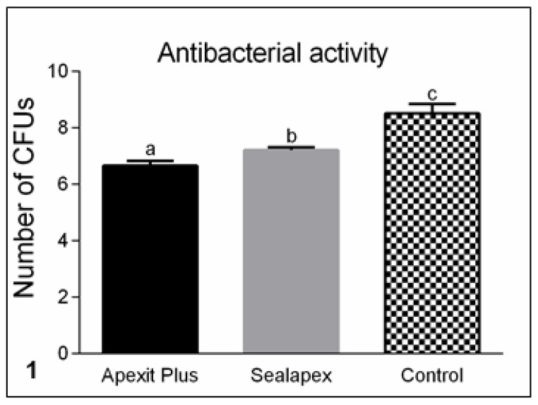
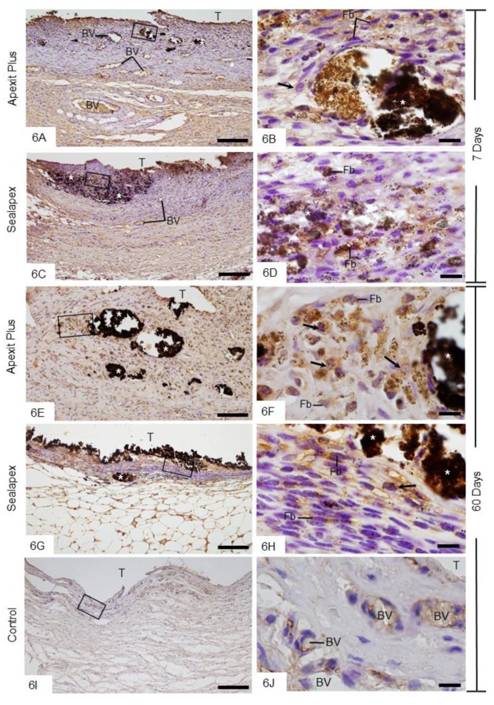
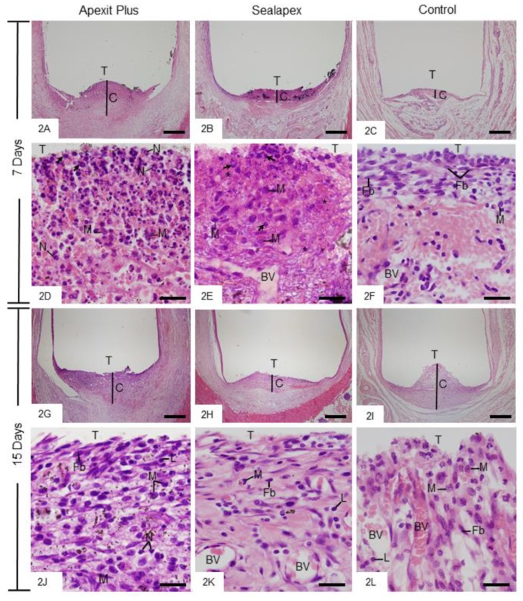
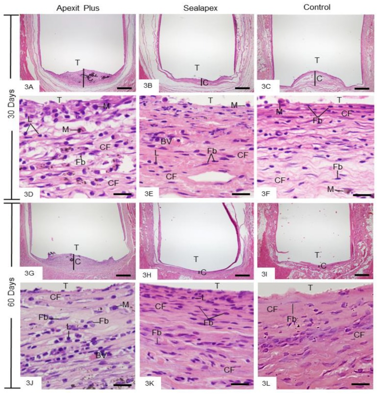
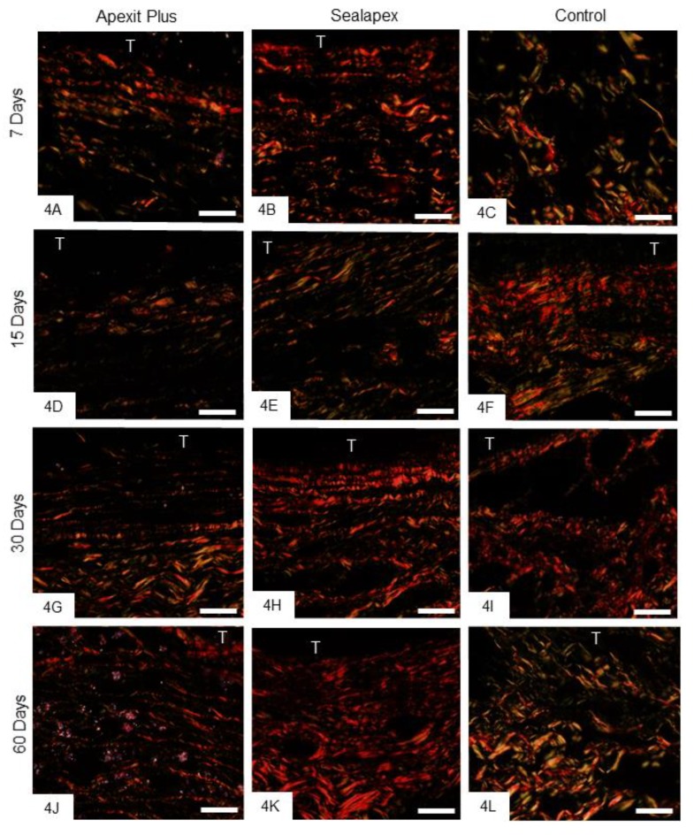
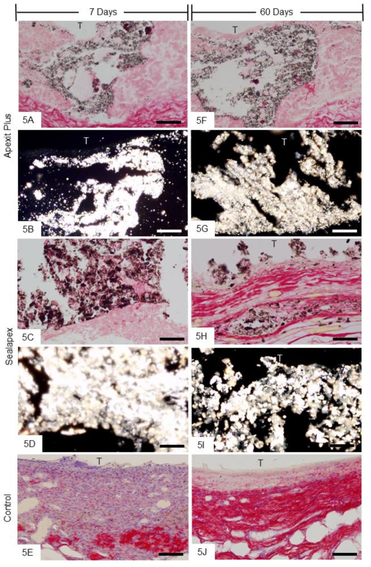
We investigated the physical properties, antimicrobial activity, and tissue reaction to Apexit Plus in comparison to Sealapex. Flow, radiopacity, setting time, and solubility were evaluated in each material. The antimicrobial activity against was performed. Polyethylene tubes containing Apexit Plus or Sealapex, and without material (control group) were implanted into the subcutaneous tissue of rats. At 7, 15, 30, and 60 days of implantation, the specimens were paraffin-embedded and the number of inflammatory cells (ICs) and the amount of birefringent collagen (BC) were quantified. The von Kossa reaction followed by immunohistochemistry for detection of alkaline phosphatase (ALP) was also performed. Statistical analysis was performed with ANOVA and Tukey test ( ≤ 0.05). The flow value of Apexit Plus was greater than Sealapex, whereas the radiopacity (3.44 mm Al) was lower than Sealapex (6.82 mm Al). Apexit Plus showed lower solubility and shorter initial and final setting ( 0.0001), whereas the antimicrobial activity was significantly greater than Sealapex. Although the number of ICs was higher in Apexit Plus ( = 0.0009) at 7 days, no significant difference was detected between Apexit Plus and Sealapex at 15, 30, and 60 days. All groups showed higher values for BC in the capsules over time. ALP-immunolabelled cells were observed, mainly around von Kossa-positive structures, either in the capsules of Apexit Plus or Sealapex. Therefore, our results revealed that Apexit Plus exhibited a greater effectiveness against and better physical properties than Sealapex, except for the radiopacity. In vivo findings indicate that Apexit Plus is biocompatible and presents potential bioactivity in the subcutaneous tissue.
我们将Apexit Plus与Sealapex进行比较,研究了其物理性能、抗菌活性及组织反应。评估了每种材料的流动性、射线不透性、凝固时间和溶解性。对其抗菌活性进行了检测。将装有Apexit Plus或Sealapex以及无材料的聚乙烯管(对照组)植入大鼠皮下组织。在植入后的第7、15、30和60天,将标本进行石蜡包埋,并对炎性细胞(ICs)数量和双折射胶原(BC)量进行定量分析。还进行了冯·科萨反应,随后进行免疫组织化学检测碱性磷酸酶(ALP)。采用方差分析和Tukey检验进行统计分析(P≤0.05)。Apexit Plus的流动值大于Sealapex,而射线不透性(3.44 mm铝当量)低于Sealapex(6.82 mm铝当量)。Apexit Plus的溶解性较低,初始和最终凝固时间较短(P<0.0001),而抗菌活性明显高于Sealapex。虽然在第7天Apexit Plus中的ICs数量较高(P = 0.0009),但在第15、30和60天Apexit Plus与Sealapex之间未检测到显著差异。随着时间推移,所有组包膜中的BC值均较高。在Apexit Plus或Sealapex的包膜中均观察到ALP免疫标记细胞,主要围绕冯·科萨阳性结构。因此,我们的结果表明,除射线不透性外,Apexit Plus对(某种细菌,原文未明确)的有效性高于Sealapex,且物理性能更好。体内研究结果表明,Apexit Plus具有生物相容性,在皮下组织中具有潜在生物活性。