Department of Biomedical Engineering and Sciences, School of Mechanical and Manufacturing Engineering, National University of Sciences and Technology, Islamabad 44000, Pakistan.
Department of Electrical and Computer Engineering, Memorial University of Newfoundland, Newfoundland, St. John's, NL A1C 5S7 P.O. Box 4200, Canada.
Sensors (Basel). 2020 Mar 13;20(6):1601. doi: 10.3390/s20061601.
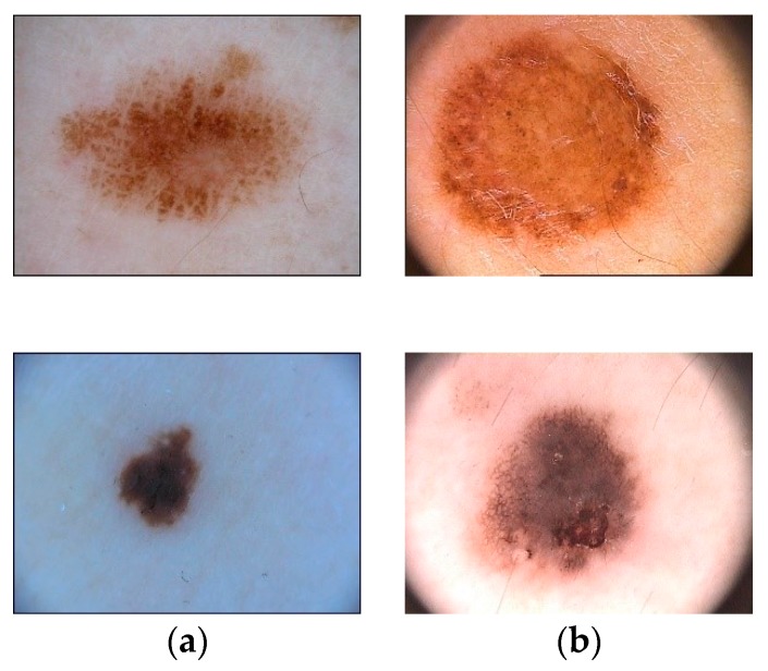
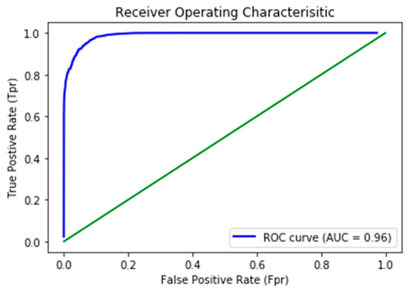
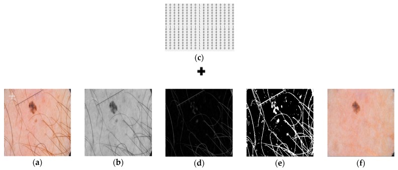
Clinical treatment of skin lesion is primarily dependent on timely detection and delimitation of lesion boundaries for accurate cancerous region localization. Prevalence of skin cancer is on the higher side, especially that of melanoma, which is aggressive in nature due to its high metastasis rate. Therefore, timely diagnosis is critical for its treatment before the onset of malignancy. To address this problem, medical imaging is used for the analysis and segmentation of lesion boundaries from dermoscopic images. Various methods have been used, ranging from visual inspection to the textural analysis of the images. However, accuracy of these methods is low for proper clinical treatment because of the sensitivity involved in surgical procedures or drug application. This presents an opportunity to develop an automated model with good accuracy so that it may be used in a clinical setting. This paper proposes an automated method for segmenting lesion boundaries that combines two architectures, the U-Net and the ResNet, collectively called Res-Unet. Moreover, we also used image inpainting for hair removal, which improved the segmentation results significantly. We trained our model on the ISIC 2017 dataset and validated it on the ISIC 2017 test set as well as the PH dataset. Our proposed model attained a Jaccard Index of 0.772 on the ISIC 2017 test set and 0.854 on the PH dataset, which are comparable results to the current available state-of-the-art techniques.
皮肤病变的临床治疗主要依赖于及时检测和划定病变边界,以准确定位癌症区域。皮肤癌的发病率较高,尤其是恶性黑色素瘤,由于其高转移率,恶性程度较高。因此,在恶变发生之前,及时诊断对于治疗至关重要。为了解决这个问题,医学成像被用于分析和分割皮肤镜图像中的病变边界。已经使用了各种方法,从目视检查到图像的纹理分析。然而,由于手术过程或药物应用的敏感性,这些方法的准确性对于正确的临床治疗来说较低。这为开发具有良好准确性的自动化模型提供了机会,以便在临床环境中使用。本文提出了一种结合 U-Net 和 ResNet 两种架构的自动分割病变边界的方法,称为 Res-Unet。此外,我们还使用图像修复进行毛发去除,这显著提高了分割结果。我们在 ISIC 2017 数据集上训练我们的模型,并在 ISIC 2017 测试集以及 PH 数据集上进行验证。我们提出的模型在 ISIC 2017 测试集上的 Jaccard 指数为 0.772,在 PH 数据集上的 Jaccard 指数为 0.854,与当前可用的最先进技术相当。