• 文献检索
  • 文档翻译
  • 深度研究
  • 学术资讯
  • Suppr Zotero 插件Zotero 插件
  • 邀请有礼
  • 套餐&价格
  • 历史记录
应用&插件
Suppr Zotero 插件Zotero 插件浏览器插件Mac 客户端Windows 客户端微信小程序
定价
高级版会员购买积分包购买API积分包
服务
文献检索文档翻译深度研究API 文档MCP 服务
关于我们
关于 Suppr公司介绍联系我们用户协议隐私条款
关注我们

Suppr 超能文献

核心技术专利:CN118964589B侵权必究
粤ICP备2023148730 号-1Suppr @ 2026

文献检索

告别复杂PubMed语法,用中文像聊天一样搜索,搜遍4000万医学文献。AI智能推荐,让科研检索更轻松。

立即免费搜索

文件翻译

保留排版,准确专业,支持PDF/Word/PPT等文件格式,支持 12+语言互译。

免费翻译文档

深度研究

AI帮你快速写综述,25分钟生成高质量综述,智能提取关键信息,辅助科研写作。

立即免费体验

使用最先进的深度学习架构进行自动视神经头定位和杯盘比检测。

Automatic optic nerve head localization and cup-to-disc ratio detection using state-of-the-art deep-learning architectures.

机构信息

Department of Ophthalmology, Pusan National University College of Medicine, Busan, Korea.

Biomedical Research Institute, Pusan National University Hospital, Busan, Korea.

出版信息

Sci Rep. 2020 Mar 19;10(1):5025. doi: 10.1038/s41598-020-62022-x.

DOI:10.1038/s41598-020-62022-x
PMID:32193499
原文链接:https://pmc.ncbi.nlm.nih.gov/articles/PMC7081256/
Abstract

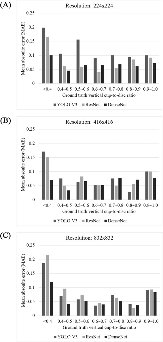
Computer vision has greatly advanced recently. Since AlexNet was first introduced, many modified deep learning architectures have been developed and they are still evolving. However, there are few studies comparing these architectures in the field of ophthalmology. This study compared the performance of various state-of-the-art deep-learning architectures for detecting the optic nerve head and vertical cup-to-disc ratio in fundus images. Three different architectures were compared: YOLO V3, ResNet, and DenseNet. We compared various aspects of performance, which were not confined to the accuracy of detection but included, as well, the processing time, diagnostic performance, effect of the graphic processing unit (GPU), and image resolution. In general, as the input image resolution increased, the classification accuracy, localization error, and diagnostic performance all improved, but the optimal architecture differed depending on the resolution. The processing time was significantly accelerated with GPU assistance; even at the high resolution of 832 × 832, it was approximately 170 ms, which was at least 26 times slower without GPU. The choice of architecture may depend on the researcher's purpose when balancing between speed and accuracy. This study provides a guideline to determine deep learning architecture, optimal image resolution, and the appropriate hardware.

摘要

计算机视觉技术最近取得了重大进展。自从 AlexNet 首次问世以来,已经开发出了许多改进的深度学习架构,并且它们仍在不断发展。然而,在眼科领域,很少有研究比较这些架构。本研究比较了各种最先进的深度学习架构在检测眼底图像中的视神经头和垂直杯盘比方面的性能。比较了三种不同的架构:YOLO V3、ResNet 和 DenseNet。我们比较了性能的各个方面,不仅限于检测的准确性,还包括处理时间、诊断性能、图形处理单元 (GPU) 的影响以及图像分辨率。一般来说,随着输入图像分辨率的增加,分类准确性、定位误差和诊断性能都得到了提高,但最佳架构取决于分辨率。GPU 辅助显著加速了处理时间;即使在 832×832 的高分辨率下,处理时间也大约为 170ms,而没有 GPU 的处理时间至少慢 26 倍。架构的选择可能取决于研究人员在速度和准确性之间的权衡目的。本研究提供了一个确定深度学习架构、最佳图像分辨率和合适硬件的指导方针。

https://cdn.ncbi.nlm.nih.gov/pmc/blobs/e3e5/7081256/f3dbd157f747/41598_2020_62022_Fig3_HTML.jpg
https://cdn.ncbi.nlm.nih.gov/pmc/blobs/e3e5/7081256/cf97f5ebab21/41598_2020_62022_Fig1_HTML.jpg
https://cdn.ncbi.nlm.nih.gov/pmc/blobs/e3e5/7081256/2d071c9c70de/41598_2020_62022_Fig2_HTML.jpg
https://cdn.ncbi.nlm.nih.gov/pmc/blobs/e3e5/7081256/f3dbd157f747/41598_2020_62022_Fig3_HTML.jpg
https://cdn.ncbi.nlm.nih.gov/pmc/blobs/e3e5/7081256/cf97f5ebab21/41598_2020_62022_Fig1_HTML.jpg
https://cdn.ncbi.nlm.nih.gov/pmc/blobs/e3e5/7081256/2d071c9c70de/41598_2020_62022_Fig2_HTML.jpg
https://cdn.ncbi.nlm.nih.gov/pmc/blobs/e3e5/7081256/f3dbd157f747/41598_2020_62022_Fig3_HTML.jpg

相似文献

1
Automatic optic nerve head localization and cup-to-disc ratio detection using state-of-the-art deep-learning architectures.使用最先进的深度学习架构进行自动视神经头定位和杯盘比检测。
Sci Rep. 2020 Mar 19;10(1):5025. doi: 10.1038/s41598-020-62022-x.
2
Fully automated method for glaucoma screening using robust optic nerve head detection and unsupervised segmentation based cup-to-disc ratio computation in retinal fundus images.基于视网膜眼底图像中稳健的视神经头检测和无监督视杯/视盘比计算的青光眼筛查全自动方法。
Comput Med Imaging Graph. 2019 Oct;77:101643. doi: 10.1016/j.compmedimag.2019.101643. Epub 2019 Aug 14.
3
WGAN domain adaptation for the joint optic disc-and-cup segmentation in fundus images.基于 WGAN 的眼底图像视盘-杯联合分割领域自适应方法。
Int J Comput Assist Radiol Surg. 2020 Jul;15(7):1205-1213. doi: 10.1007/s11548-020-02144-9. Epub 2020 May 22.
4
Optic Disc and Cup Segmentation with Blood Vessel Removal from Fundus Images for Glaucoma Detection.用于青光眼检测的眼底图像视盘和视杯分割及血管去除
Annu Int Conf IEEE Eng Med Biol Soc. 2018 Jul;2018:862-865. doi: 10.1109/EMBC.2018.8512400.
5
Accurate, fast, data efficient and interpretable glaucoma diagnosis with automated spatial analysis of the whole cup to disc profile.利用全自动的视杯-视盘整体形态分析实现准确、快速、高效的数据利用和可解释的青光眼诊断。
PLoS One. 2019 Jan 10;14(1):e0209409. doi: 10.1371/journal.pone.0209409. eCollection 2019.
6
Diagnosis of Glaucoma on Retinal Fundus Images Using Deep Learning: Detection of Nerve Fiber Layer Defect and Optic Disc Analysis.基于深度学习的眼底图像青光眼诊断:神经纤维层缺损检测和视盘分析。
Adv Exp Med Biol. 2020;1213:121-132. doi: 10.1007/978-3-030-33128-3_8.
7
The region of interest localization for glaucoma analysis from retinal fundus image using deep learning.利用深度学习进行视网膜眼底图像的青光眼分析的感兴趣区域定位。
Comput Methods Programs Biomed. 2018 Oct;165:25-35. doi: 10.1016/j.cmpb.2018.08.003. Epub 2018 Aug 8.
8
Computer-aided diagnosis of glaucoma using fundus images: A review.基于眼底图像的青光眼计算机辅助诊断:综述
Comput Methods Programs Biomed. 2018 Oct;165:1-12. doi: 10.1016/j.cmpb.2018.07.012. Epub 2018 Jul 26.
9
Deep learning on fundus images detects glaucoma beyond the optic disc.眼底图像深度学习可在视盘之外检测青光眼。
Sci Rep. 2021 Oct 13;11(1):20313. doi: 10.1038/s41598-021-99605-1.
10
Efficacy of a Deep Learning System for Detecting Glaucomatous Optic Neuropathy Based on Color Fundus Photographs.基于眼底彩色照片的深度学习系统检测青光眼视神经病变的效果。
Ophthalmology. 2018 Aug;125(8):1199-1206. doi: 10.1016/j.ophtha.2018.01.023. Epub 2018 Mar 2.

引用本文的文献

1
Independent Evaluation of RETFound Foundation Model's Performance on Optic Nerve Analysis Using Fundus Photography.关于使用眼底摄影术对RETFound基础模型在视神经分析方面的性能进行独立评估
Ophthalmol Sci. 2025 Jan 28;5(3):100720. doi: 10.1016/j.xops.2025.100720. eCollection 2025 May-Jun.
2
Diabetic Retinopathy Features Segmentation without Coding Experience with Computer Vision Models YOLOv8 and YOLOv9.无需编码经验,使用计算机视觉模型YOLOv8和YOLOv9对糖尿病视网膜病变特征进行分割
Vision (Basel). 2024 Aug 23;8(3):48. doi: 10.3390/vision8030048.
3
Automated vertical cup-to-disc ratio determination from fundus images for glaucoma detection.
自动化眼底图像杯盘比测量在青光眼检测中的应用。
Sci Rep. 2024 Feb 24;14(1):4494. doi: 10.1038/s41598-024-55056-y.
4
Superpixel-Based Optic Nerve Head Segmentation Method of Fundus Images for Glaucoma Assessment.基于超像素的用于青光眼评估的眼底图像视盘分割方法
Diagnostics (Basel). 2022 Dec 17;12(12):3210. doi: 10.3390/diagnostics12123210.
5
The use of deep learning technology for the detection of optic neuropathy.深度学习技术在视神经病变检测中的应用。
Quant Imaging Med Surg. 2022 Mar;12(3):2129-2143. doi: 10.21037/qims-21-728.
6
Application of Artificial Intelligence and Deep Learning for Choroid Segmentation in Myopia.人工智能和深度学习在近视脉络膜分割中的应用。
Transl Vis Sci Technol. 2022 Feb 1;11(2):38. doi: 10.1167/tvst.11.2.38.
7
Automatic Detection of Coronary Metallic Stent Struts Based on YOLOv3 and R-FCN.基于 YOLOv3 和 R-FCN 的冠状动脉金属支架梁的自动检测。
Comput Math Methods Med. 2020 Sep 1;2020:1793517. doi: 10.1155/2020/1793517. eCollection 2020.