Department of Eye & Vision Science, Institute of Ageing and Chronic Disease, University of Liverpool, Liverpool, United Kingdom.
Centre for Clinical Brain Sciences, University of Edinburgh, Chancellor's Building, Edinburgh, United Kingdom.
PLoS One. 2019 Jan 10;14(1):e0209409. doi: 10.1371/journal.pone.0209409. eCollection 2019.
Glaucoma is the leading cause of irreversible blindness worldwide. It is a heterogeneous group of conditions with a common optic neuropathy and associated loss of peripheral vision. Both over and under-diagnosis carry high costs in terms of healthcare spending and preventable blindness. The characteristic clinical feature of glaucoma is asymmetrical optic nerve rim narrowing, which is difficult for humans to quantify reliably. Strategies to improve and automate optic disc assessment are therefore needed to prevent sight loss.
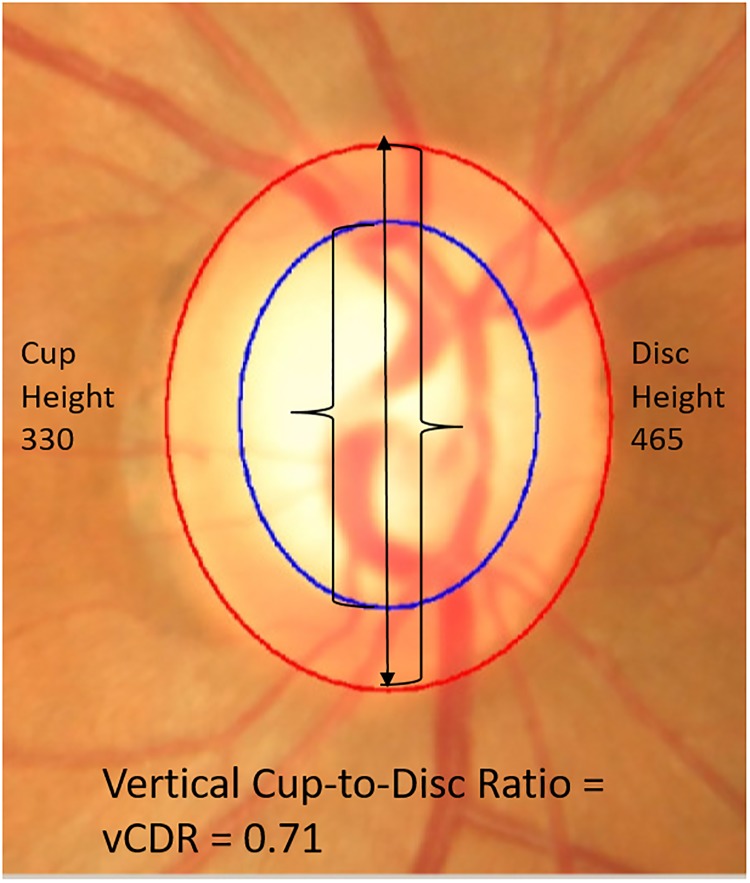
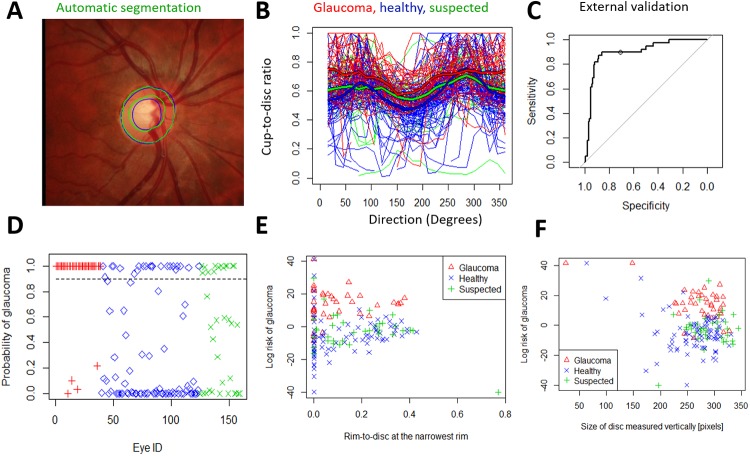
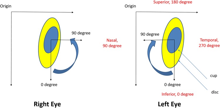
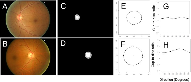
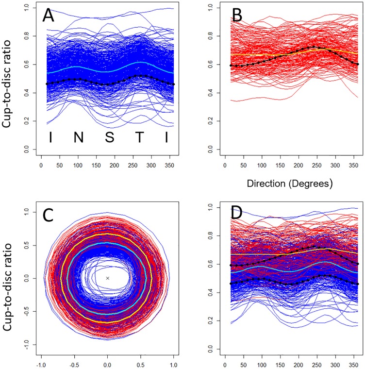
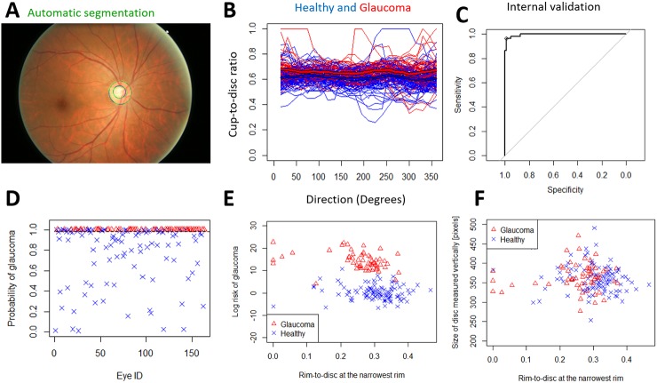
We developed a novel glaucoma detection algorithm that segments and analyses colour photographs to quantify optic nerve rim consistency around the whole disc at 15-degree intervals. This provides a profile of the cup/disc ratio, in contrast to the vertical cup/disc ratio in common use. We introduce a spatial probabilistic model, to account for the optic nerve shape, we then use this model to derive a disc deformation index and a decision rule for glaucoma. We tested our algorithm on two separate image datasets (ORIGA and RIM-ONE).
The spatial algorithm accurately distinguished glaucomatous and healthy discs on internal and external validation (AUROC 99.6% and 91.0% respectively). It achieves this using a dataset 100-times smaller than that required for deep learning algorithms, is flexible to the type of cup and disc segmentation (automated or semi-automated), utilises images with missing data, and is correlated with the disc size (p = 0.02) and the rim-to-disc at the narrowest rim (p<0.001, in external validation).
The spatial probabilistic algorithm is highly accurate, highly data efficient and it extends to any imaging hardware in which the boundaries of cup and disc can be segmented, thus making the algorithm particularly applicable to research into disease mechanisms, and also glaucoma screening in low resource settings.
青光眼是全球导致不可逆性失明的主要原因。它是一组具有共同视神经病变和相关周边视力丧失的异质性疾病。过度和诊断不足都会导致医疗保健支出增加和可预防失明。青光眼的特征性临床特征是视神经边缘不对称性变窄,这很难让人类可靠地进行定量。因此,需要改进和自动化视神经盘评估策略,以防止视力丧失。
我们开发了一种新的青光眼检测算法,该算法通过分段和分析彩色照片,以 15 度间隔量化整个视盘周围的视神经边缘一致性。这提供了杯/盘比的轮廓,与常用的垂直杯/盘比形成对比。我们引入了一种空间概率模型,以解释视神经的形状,然后使用该模型得出一个盘变形指数和用于诊断青光眼的决策规则。我们在两个独立的图像数据集(ORIGA 和 RIM-ONE)上测试了我们的算法。
空间算法在内部和外部验证中准确地区分了青光眼和健康的视盘(AUROC 分别为 99.6%和 91.0%)。它使用比深度学习算法所需的数据集小 100 倍的数据集实现了这一点,对杯和盘分割的类型具有灵活性(自动或半自动),利用具有缺失数据的图像,并且与视盘大小相关(p=0.02)和最窄边缘处的边缘到盘(p<0.001,在外部验证中)。
空间概率算法具有高度准确性、高效的数据利用能力,并且可以扩展到任何可以分割杯和盘边界的成像硬件,因此特别适用于疾病机制的研究,也适用于资源匮乏环境中的青光眼筛查。