Simons Centre for the Study of Living Machines, National Centre for Biological Sciences (TIFR), Bangalore 560065, India.
National Centre for Biological Sciences (TIFR), Bangalore 560065, India.
Sci Adv. 2020 Mar 11;6(11):eaay6093. doi: 10.1126/sciadv.aay6093. eCollection 2020 Mar.
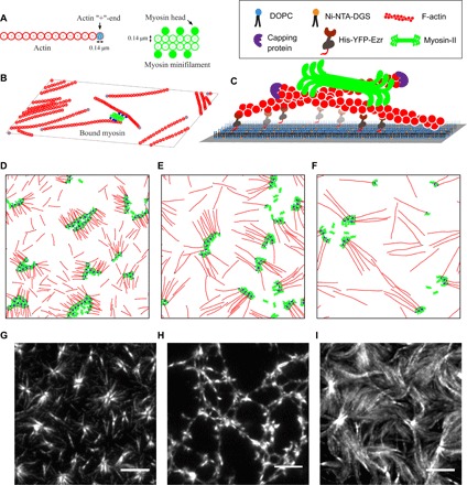
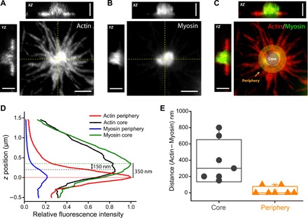
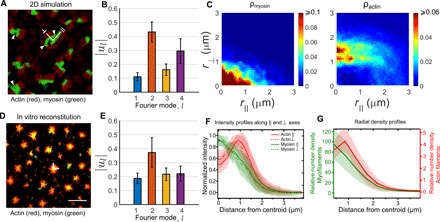
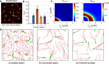
Recent in vivo studies reveal that several membrane proteins are driven to form nanoclusters by active contractile flows arising from localized dynamic patterning of F-actin and myosin at the cortex. Since myosin-II assemble as minifilaments with tens of myosin heads, one might worry that steric considerations would obstruct the emergence of nanoclustering. Using coarse-grained, agent-based simulations that account for steric constraints, we find that the patterns exhibited by actomyosin in two dimensions, do not resemble the steady-state patterns in our in vitro reconstitution of actomyosin on a supported bilayer. We perform simulations in a thin rectangular slab, separating the layer of actin filaments from myosin-II minifilaments. This recapitulates the observed features of in vitro patterning. Using super resolution microscopy, we find evidence for such stratification in our in vitro system. Our study suggests that molecular stratification may be an important organizing feature of the cortical cytoskeleton in vivo.
最近的体内研究表明,由于皮层中 F-肌动蛋白和肌球蛋白的局部动态模式,几种膜蛋白被活性收缩流驱动形成纳米簇。由于肌球蛋白 II 组装成带有数十个肌球蛋白头部的微丝,人们可能会担心空间位阻会阻碍纳米簇的出现。我们使用考虑到空间位阻的粗粒化、基于代理的模拟,发现肌动球蛋白在二维上表现出的模式与我们在体外肌球蛋白在支撑双层上的重组的稳态模式并不相似。我们在薄矩形片层中进行模拟,将肌动蛋白丝层与肌球蛋白 II 微丝分开。这再现了体外模式化的观察特征。使用超分辨率显微镜,我们在我们的体外系统中发现了这种分层的证据。我们的研究表明,分子分层可能是体内皮质细胞骨架的一个重要组织特征。