• 文献检索
  • 文档翻译
  • 深度研究
  • 学术资讯
  • Suppr Zotero 插件Zotero 插件
  • 邀请有礼
  • 套餐&价格
  • 历史记录
应用&插件
Suppr Zotero 插件Zotero 插件浏览器插件Mac 客户端Windows 客户端微信小程序
定价
高级版会员购买积分包购买API积分包
服务
文献检索文档翻译深度研究API 文档MCP 服务
关于我们
关于 Suppr公司介绍联系我们用户协议隐私条款
关注我们

Suppr 超能文献

核心技术专利:CN118964589B侵权必究
粤ICP备2023148730 号-1Suppr @ 2026

文献检索

告别复杂PubMed语法,用中文像聊天一样搜索,搜遍4000万医学文献。AI智能推荐,让科研检索更轻松。

立即免费搜索

文件翻译

保留排版,准确专业,支持PDF/Word/PPT等文件格式,支持 12+语言互译。

免费翻译文档

深度研究

AI帮你快速写综述,25分钟生成高质量综述,智能提取关键信息,辅助科研写作。

立即免费体验

用飞秒 X 射线激光捕获的光致变色蛋白的主要结构光响应。

The primary structural photoresponse of phytochrome proteins captured by a femtosecond X-ray laser.

机构信息

Department of Chemistry and Molecular Biology, University of Gothenburg, Gothenburg, Sweden.

Department of Biological and Environmental Science, Nanoscience Center, University of Jyvaskyla, Jyvaskyla, Finland.

出版信息

Elife. 2020 Mar 31;9:e53514. doi: 10.7554/eLife.53514.

DOI:10.7554/eLife.53514
PMID:32228856
原文链接:https://pmc.ncbi.nlm.nih.gov/articles/PMC7164956/
Abstract

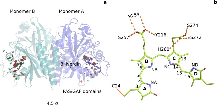
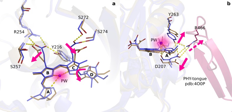
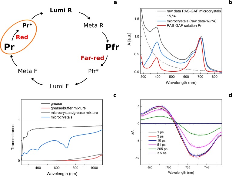
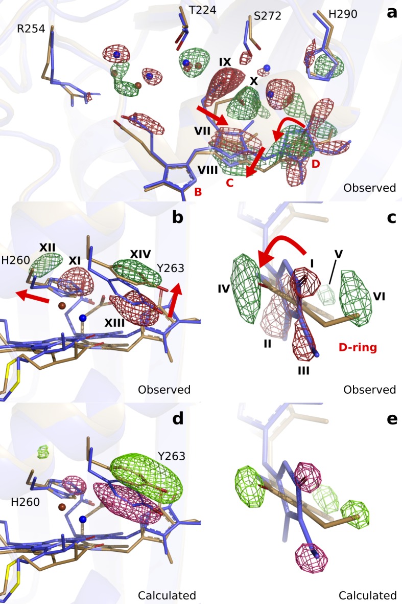
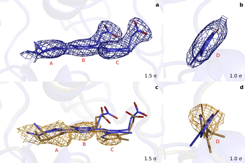
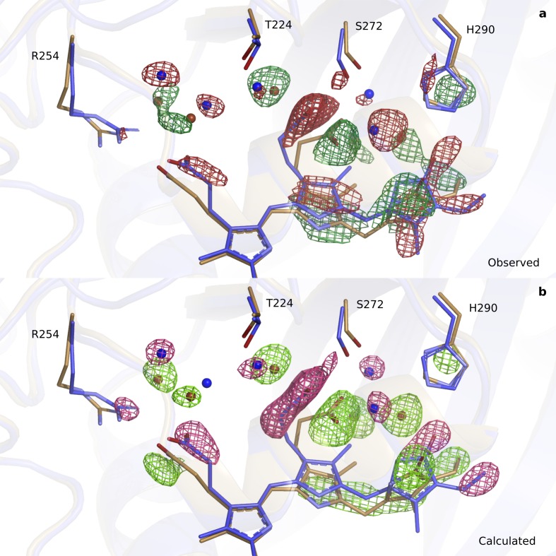
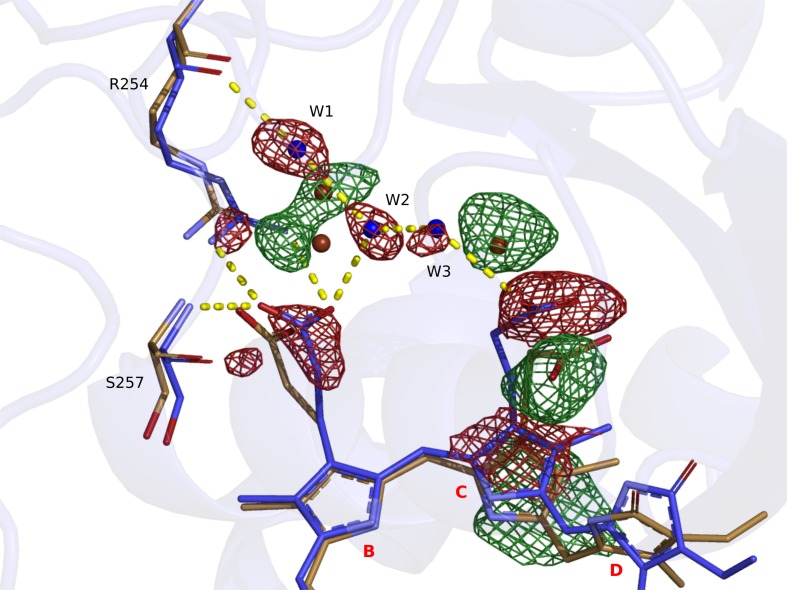
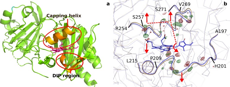
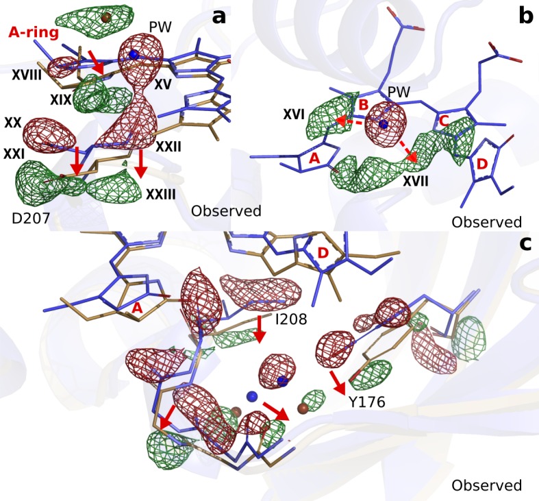
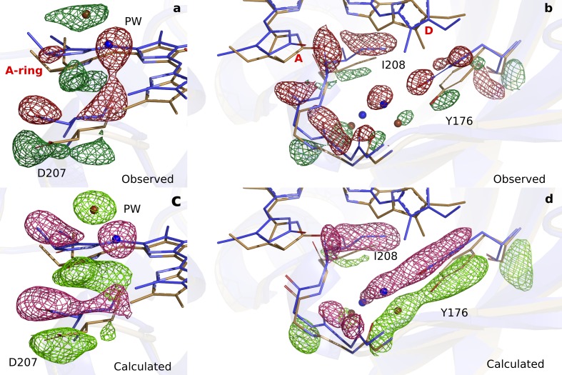
Phytochrome proteins control the growth, reproduction, and photosynthesis of plants, fungi, and bacteria. Light is detected by a bilin cofactor, but it remains elusive how this leads to activation of the protein through structural changes. We present serial femtosecond X-ray crystallographic data of the chromophore-binding domains of a bacterial phytochrome at delay times of 1 ps and 10 ps after photoexcitation. The data reveal a twist of the D-ring, which leads to partial detachment of the chromophore from the protein. Unexpectedly, the conserved so-called pyrrole water is photodissociated from the chromophore, concomitant with movement of the A-ring and a key signaling aspartate. The changes are wired together by ultrafast backbone and water movements around the chromophore, channeling them into signal transduction towards the output domains. We suggest that the observed collective changes are important for the phytochrome photoresponse, explaining the earliest steps of how plants, fungi and bacteria sense red light.

摘要

光敏色素蛋白控制着植物、真菌和细菌的生长、繁殖和光合作用。光由双吡咯辅因子检测,但目前仍不清楚这如何通过结构变化导致蛋白激活。我们呈现了细菌光敏色素的发色团结合域在光激发后 1 ps 和 10 ps 的连续飞秒 X 射线晶体学数据。数据显示 D 环的扭曲,导致发色团部分从蛋白上脱离。出乎意料的是,保守的所谓吡咯水从发色团上光解,伴随着 A 环和关键信号天冬氨酸的移动。这些变化通过发色团周围超快的骨架和水的运动紧密相连,将它们引导到信号转导到输出域。我们认为观察到的集体变化对视黄醛光反应很重要,解释了植物、真菌和细菌如何感知红光的最早步骤。

https://cdn.ncbi.nlm.nih.gov/pmc/blobs/3770/7164956/922a8baef419/elife-53514-fig5.jpg
https://cdn.ncbi.nlm.nih.gov/pmc/blobs/3770/7164956/b7dcbe0cbf80/elife-53514-fig1.jpg
https://cdn.ncbi.nlm.nih.gov/pmc/blobs/3770/7164956/ccc0703541cb/elife-53514-fig1-figsupp1.jpg
https://cdn.ncbi.nlm.nih.gov/pmc/blobs/3770/7164956/06619d37aa59/elife-53514-fig1-figsupp2.jpg
https://cdn.ncbi.nlm.nih.gov/pmc/blobs/3770/7164956/953227e34deb/elife-53514-fig1-figsupp3.jpg
https://cdn.ncbi.nlm.nih.gov/pmc/blobs/3770/7164956/0b61b5950143/elife-53514-fig1-figsupp4.jpg
https://cdn.ncbi.nlm.nih.gov/pmc/blobs/3770/7164956/ed223eec47f1/elife-53514-fig1-figsupp5.jpg
https://cdn.ncbi.nlm.nih.gov/pmc/blobs/3770/7164956/10918a5d0b77/elife-53514-fig2.jpg
https://cdn.ncbi.nlm.nih.gov/pmc/blobs/3770/7164956/5ea3d5166065/elife-53514-fig3.jpg
https://cdn.ncbi.nlm.nih.gov/pmc/blobs/3770/7164956/a22100434ba6/elife-53514-fig3-figsupp1.jpg
https://cdn.ncbi.nlm.nih.gov/pmc/blobs/3770/7164956/6eb330a864dd/elife-53514-fig3-figsupp2.jpg
https://cdn.ncbi.nlm.nih.gov/pmc/blobs/3770/7164956/cfee6135b7ac/elife-53514-fig3-figsupp3.jpg
https://cdn.ncbi.nlm.nih.gov/pmc/blobs/3770/7164956/f9944f2da715/elife-53514-fig3-figsupp4.jpg
https://cdn.ncbi.nlm.nih.gov/pmc/blobs/3770/7164956/383a0ac2ce22/elife-53514-fig4.jpg
https://cdn.ncbi.nlm.nih.gov/pmc/blobs/3770/7164956/e79af3e394d2/elife-53514-fig4-figsupp1.jpg
https://cdn.ncbi.nlm.nih.gov/pmc/blobs/3770/7164956/922a8baef419/elife-53514-fig5.jpg

相似文献

1
The primary structural photoresponse of phytochrome proteins captured by a femtosecond X-ray laser.用飞秒 X 射线激光捕获的光致变色蛋白的主要结构光响应。
Elife. 2020 Mar 31;9:e53514. doi: 10.7554/eLife.53514.
2
On the (un)coupling of the chromophore, tongue interactions, and overall conformation in a bacterial phytochrome.在细菌光致变色中的发色团、舌相互作用和整体构象的(去)偶联。
J Biol Chem. 2018 May 25;293(21):8161-8172. doi: 10.1074/jbc.RA118.001794. Epub 2018 Apr 5.
3
Crystallographic and electron microscopic analyses of a bacterial phytochrome reveal local and global rearrangements during photoconversion.对一种细菌光敏色素的晶体学和电子显微镜分析揭示了光转换过程中的局部和整体重排。
J Biol Chem. 2014 Aug 29;289(35):24573-87. doi: 10.1074/jbc.M114.571661. Epub 2014 Jul 8.
4
The room temperature crystal structure of a bacterial phytochrome determined by serial femtosecond crystallography.利用连续飞秒晶体学技术解析的细菌光光解酶的室温晶体结构。
Sci Rep. 2016 Oct 19;6:35279. doi: 10.1038/srep35279.
5
Coordination of the biliverdin D-ring in bacteriophytochromes.细菌菌视紫红质中二吡咯环 D 的协调作用。
Phys Chem Chem Phys. 2018 Jul 11;20(27):18216-18225. doi: 10.1039/c8cp01696h.
6
Signal amplification and transduction in phytochrome photosensors.光敏色素光传感器中的信号放大和转导。
Nature. 2014 May 8;509(7499):245-248. doi: 10.1038/nature13310. Epub 2014 Apr 30.
7
Signaling Mechanism of Phytochromes in Solution.植物色素在溶液中的信号机制。
Structure. 2021 Feb 4;29(2):151-160.e3. doi: 10.1016/j.str.2020.08.009. Epub 2020 Sep 10.
8
Chromophore-Protein Interplay during the Phytochrome Photocycle Revealed by Step-Scan FTIR Spectroscopy.利用分步扫描傅里叶变换红外光谱技术揭示光色素光循环过程中的生色团-蛋白质相互作用。
J Am Chem Soc. 2018 Oct 3;140(39):12396-12404. doi: 10.1021/jacs.8b04659. Epub 2018 Sep 18.
9
High-resolution crystal structures of transient intermediates in the phytochrome photocycle.光致变色循环中瞬态中间产物的高分辨率晶体结构。
Structure. 2021 Jul 1;29(7):743-754.e4. doi: 10.1016/j.str.2021.03.004. Epub 2021 Mar 22.
10
X-ray radiation induces deprotonation of the bilin chromophore in crystalline D. radiodurans phytochrome.X射线辐射会诱导嗜放射栖热菌光敏色素晶体中的胆色素发色团发生去质子化。
J Am Chem Soc. 2015 Mar 4;137(8):2792-5. doi: 10.1021/ja510923m. Epub 2015 Feb 18.

引用本文的文献

1
Engineered bacteriophytochrome heterodimers for research and applications.用于研究和应用的工程化细菌光敏色素异二聚体
J Biol Chem. 2025 Jul 4;301(8):110452. doi: 10.1016/j.jbc.2025.110452.
2
Pr and Pfr structures of plant phytochrome A.植物光敏色素A的红光吸收型和远红光吸收型结构
Nat Commun. 2025 Jun 21;16(1):5319. doi: 10.1038/s41467-025-60738-w.
3
Distinct Protochromic Mechanisms Driving Green/Red Absorption in Phycocyanobilin-Binding Proteins.驱动藻胆青素结合蛋白中绿色/红色吸收的不同原色机制。

本文引用的文献

1
Modulation of Structural Heterogeneity Controls Phytochrome Photoswitching.结构异质性的调控控制着光敏色素的光开关。
Biophys J. 2020 Jan 21;118(2):415-421. doi: 10.1016/j.bpj.2019.11.025. Epub 2019 Nov 26.
2
Time-resolved serial femtosecond crystallography at the European XFEL.时间分辨连续飞秒晶体学在欧洲 X 射线自由电子激光装置上的应用。
Nat Methods. 2020 Jan;17(1):73-78. doi: 10.1038/s41592-019-0628-z. Epub 2019 Nov 18.
3
High-resolution crystal structures of a myxobacterial phytochrome at cryo and room temperatures.一种粘细菌光敏色素在低温和室温下的高分辨率晶体结构。
Biochemistry. 2025 Jul 1;64(13):2823-2833. doi: 10.1021/acs.biochem.4c00870. Epub 2025 Jun 18.
4
Observation of early events in the photoactivation of Myxobacterial phytochrome using time-resolved serial femtosecond crystallography.利用时间分辨串联飞秒晶体学观察粘细菌光敏色素光激活的早期事件。
Commun Chem. 2025 Jun 12;8(1):183. doi: 10.1038/s42004-025-01578-z.
5
Direct visualization of electric-field-stimulated ion conduction in a potassium channel.钾通道中电场刺激离子传导的直接可视化
Cell. 2025 Jan 9;188(1):77-88.e15. doi: 10.1016/j.cell.2024.12.006.
6
Structural effects of high laser power densities on an early bacteriorhodopsin photocycle intermediate.高激光功率密度对早期菌紫质光循环中间产物的结构影响。
Nat Commun. 2024 Nov 27;15(1):10278. doi: 10.1038/s41467-024-54422-8.
7
ResiDEM: Analytical Tool for Isomorphous Difference Electron Density Maps Utilizing Dynamic Residue Identification via Density Clustering.ResiDEM:利用密度聚类进行动态残基识别的同晶差电子密度图分析工具。
J Chem Inf Model. 2024 Oct 14;64(19):7565-7575. doi: 10.1021/acs.jcim.4c00858. Epub 2024 Sep 19.
8
Molecular origins of absorption wavelength variation among phycocyanobilin-binding proteins.藻蓝胆素结合蛋白吸收波长变化的分子起源
Biophys J. 2024 Oct 1;123(19):3375-3385. doi: 10.1016/j.bpj.2024.08.001. Epub 2024 Aug 8.
9
Analogies and Differences in the Photoactivation Mechanism of Bathy and Canonical Bacteriophytochromes Revealed by Multiscale Modeling.多尺度建模揭示的深海细菌光敏色素与典型细菌光敏色素光激活机制的类比与差异
J Phys Chem Lett. 2024 Aug 8;15(31):8078-8084. doi: 10.1021/acs.jpclett.4c01823. Epub 2024 Aug 1.
10
Appraising protein conformational changes by resampling time-resolved serial x-ray crystallography data.通过重采样时间分辨串行X射线晶体学数据评估蛋白质构象变化。
Struct Dyn. 2024 Jul 24;11(4):044302. doi: 10.1063/4.0000258. eCollection 2024 Jul.
Struct Dyn. 2019 Sep 17;6(5):054701. doi: 10.1063/1.5120527. eCollection 2019 Sep.
4
Structural snapshot of a bacterial phytochrome in its functional intermediate state.细菌光致变色蛋白功能中间态的结构快照。
Nat Commun. 2018 Nov 21;9(1):4912. doi: 10.1038/s41467-018-07392-7.
5
Chromophore-Protein Interplay during the Phytochrome Photocycle Revealed by Step-Scan FTIR Spectroscopy.利用分步扫描傅里叶变换红外光谱技术揭示光色素光循环过程中的生色团-蛋白质相互作用。
J Am Chem Soc. 2018 Oct 3;140(39):12396-12404. doi: 10.1021/jacs.8b04659. Epub 2018 Sep 18.
6
Retinal isomerization in bacteriorhodopsin captured by a femtosecond x-ray laser.利用飞秒 X 射线激光捕获的菌紫质中的视网膜异构化。
Science. 2018 Jul 13;361(6398). doi: 10.1126/science.aat0094. Epub 2018 Jun 14.
7
3D Structures of Plant Phytochrome A as Pr and Pfr From Solid-State NMR: Implications for Molecular Function.基于固态核磁共振的植物光敏色素A Pr和Pfr的3D结构:对分子功能的启示
Front Plant Sci. 2018 Apr 24;9:498. doi: 10.3389/fpls.2018.00498. eCollection 2018.
8
Correlating structural and photochemical heterogeneity in cyanobacteriochrome NpR6012g4.关联结构和光化学异质性在蓝藻菌视紫红质 NpR6012g4 中。
Proc Natl Acad Sci U S A. 2018 Apr 24;115(17):4387-4392. doi: 10.1073/pnas.1720682115. Epub 2018 Apr 9.
9
Chromophore twisting in the excited state of a photoswitchable fluorescent protein captured by time-resolved serial femtosecond crystallography.通过时间分辨连续飞秒晶体学捕获的光致变色荧光蛋白激发态中的生色团扭曲。
Nat Chem. 2018 Jan;10(1):31-37. doi: 10.1038/nchem.2853. Epub 2017 Sep 11.
10
The room temperature crystal structure of a bacterial phytochrome determined by serial femtosecond crystallography.利用连续飞秒晶体学技术解析的细菌光光解酶的室温晶体结构。
Sci Rep. 2016 Oct 19;6:35279. doi: 10.1038/srep35279.