Hunan Provincial Key Laboratory of Intelligent Computing and Language Information Processing, Hunan Normal University, Changsha, Hunan, 410081, China.
School of Automation, Central South University, Changsha, Hunan, 410083, China.
Sci Rep. 2020 Apr 10;10(1):6256. doi: 10.1038/s41598-020-63242-x.
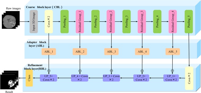
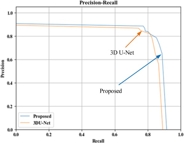
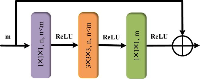
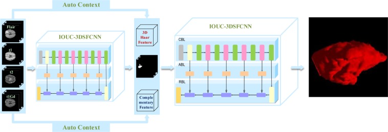
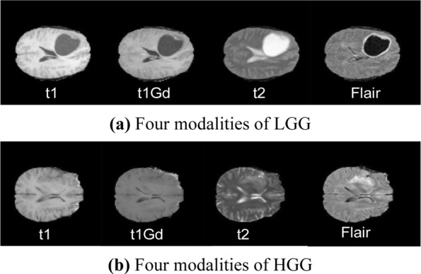
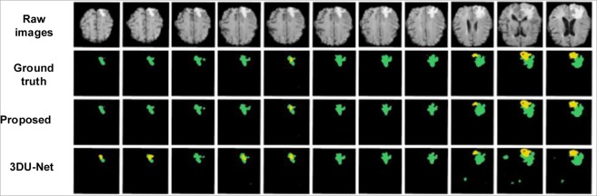
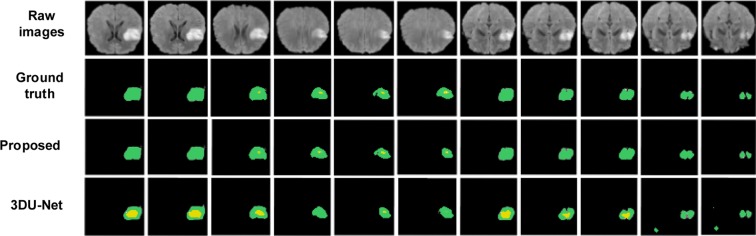
Accurate segmentation of brain tumors from magnetic resonance (MR) images play a pivot role in assisting diagnoses, treatments and postoperative evaluations. However, due to its structural complexities, e.g., fuzzy tumor boundaries with irregular shapes, accurate 3D brain tumor delineation is challenging. In this paper, an intersection over union (IOU) constraint 3D symmetric full convolutional neural network (IOUC-3DSFCNN) model fused with multimodal auto-context is proposed for the 3D brain tumor segmentation. IOUC-3DSFCNN incorporates 3D residual groups into the classic 3DU-Net to further deepen the network structure to obtain more abstract voxel features under a five-layer cohesion architecture to ensure the model stability. The IOU constraint is used to address the issue of extremely unbalanced tumor foreground and background regions in MR images. In addition, to obtain more comprehensive and stable 3D brain tumor profiles, the multimodal auto-context information is fused into the IOUC-3DSFCNN model to achieve end-to-end 3D brain tumor profiles. Extensive confirmatory and comparative experiments conducted on the benchmark BRATS 2017 dataset demonstrate that the proposed segmentation model is superior to classic 3DU-Net-relevant and other state-of-the-art segmentation models, which can achieve accurate 3D tumor profiles on multimodal MRI volumes even with blurred tumor boundaries and big noise.
从磁共振(MR)图像中准确分割脑肿瘤在辅助诊断、治疗和术后评估中起着关键作用。然而,由于其结构复杂,例如,肿瘤边界模糊且形状不规则,因此准确的 3D 脑肿瘤勾画具有挑战性。在本文中,提出了一种结合多模态自上下文的交并比(IOU)约束 3D 对称全卷积神经网络(IOUC-3DSFCNN)模型,用于 3D 脑肿瘤分割。IOUC-3DSFCNN 将 3D 残差组融入到经典的 3DU-Net 中,进一步加深网络结构,在五层凝聚架构下获得更多抽象的体素特征,以确保模型稳定性。IOU 约束用于解决 MR 图像中肿瘤前景和背景区域极度不平衡的问题。此外,为了获得更全面和稳定的 3D 脑肿瘤轮廓,将多模态自上下文信息融合到 IOUC-3DSFCNN 模型中,以实现端到端的 3D 脑肿瘤轮廓。在基准 BRATS 2017 数据集上进行的广泛验证和对比实验表明,所提出的分割模型优于经典的 3DU-Net 相关模型和其他最先进的分割模型,即使肿瘤边界模糊且存在较大噪声,也可以在多模态 MRI 容积上实现准确的 3D 肿瘤轮廓。