• 文献检索
  • 文档翻译
  • 深度研究
  • 学术资讯
  • Suppr Zotero 插件Zotero 插件
  • 邀请有礼
  • 套餐&价格
  • 历史记录
应用&插件
Suppr Zotero 插件Zotero 插件浏览器插件Mac 客户端Windows 客户端微信小程序
定价
高级版会员购买积分包购买API积分包
服务
文献检索文档翻译深度研究API 文档MCP 服务
关于我们
关于 Suppr公司介绍联系我们用户协议隐私条款
关注我们

Suppr 超能文献

核心技术专利:CN118964589B侵权必究
粤ICP备2023148730 号-1Suppr @ 2026

文献检索

告别复杂PubMed语法,用中文像聊天一样搜索,搜遍4000万医学文献。AI智能推荐,让科研检索更轻松。

立即免费搜索

文件翻译

保留排版,准确专业,支持PDF/Word/PPT等文件格式,支持 12+语言互译。

免费翻译文档

深度研究

AI帮你快速写综述,25分钟生成高质量综述,智能提取关键信息,辅助科研写作。

立即免费体验

Znet:二维 MRI 脑肿瘤分割的深度学习方法。

Znet: Deep Learning Approach for 2D MRI Brain Tumor Segmentation.

机构信息

Department of Information SystemsYarmouk University Irbid 21163 Jordan.

Statistics Online Computational ResourceDepartments of Health Behavior and Biological Sciences and Computational Medicine and BioinformaticsUniversity of Michigan Ann Arbor MI 48109 USA.

出版信息

IEEE J Transl Eng Health Med. 2022 May 23;10:1800508. doi: 10.1109/JTEHM.2022.3176737. eCollection 2022.

DOI:10.1109/JTEHM.2022.3176737
PMID:35774412
原文链接:https://pmc.ncbi.nlm.nih.gov/articles/PMC9236306/
Abstract

BACKGROUND

Detection and segmentation of brain tumors using MR images are challenging and valuable tasks in the medical field. Early diagnosing and localizing of brain tumors can save lives and provide timely options for physicians to select efficient treatment plans. Deep learning approaches have attracted researchers in medical imaging due to their capacity, performance, and potential to assist in accurate diagnosis, prognosis, and medical treatment technologies.

METHODS AND PROCEDURES

This paper presents a novel framework for segmenting 2D brain tumors in MR images using deep neural networks (DNN) and utilizing data augmentation strategies. The proposed approach (Znet) is based on the idea of skip-connection, encoder-decoder architectures, and data amplification to propagate the intrinsic affinities of a relatively smaller number of expert delineated tumors, e.g., hundreds of patients of the low-grade glioma (LGG), to many thousands of synthetic cases.

RESULTS

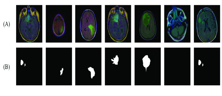
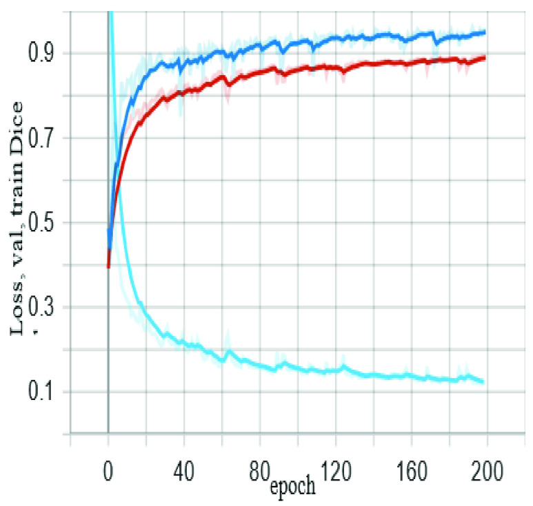
Our experimental results showed high values of the mean dice similarity coefficient (dice = 0.96 during model training and dice = 0.92 for the independent testing dataset). Other evaluation measures were also relatively high, e.g., pixel accuracy = 0.996, F1 score = 0.81, and Matthews Correlation Coefficient, MCC = 0.81. The results and visualization of the DNN-derived tumor masks in the testing dataset showcase the ZNet model's capability to localize and auto-segment brain tumors in MR images. This approach can further be generalized to 3D brain volumes, other pathologies, and a wide range of image modalities.

CONCLUSION

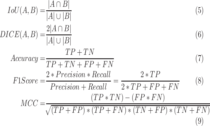
We can confirm the ability of deep learning methods and the proposed Znet framework to detect and segment tumors in MR images. Furthermore, pixel accuracy evaluation may not be a suitable evaluation measure for semantic segmentation in case of class imbalance in MR images segmentation. This is because the dominant class in ground truth images is the background. Therefore, a high value of pixel accuracy can be misleading in some computer vision applications. On the other hand, alternative evaluation metrics, such as dice and IoU (Intersection over Union), are more factual for semantic segmentation.

CLINICAL IMPACT

Artificial intelligence (AI) applications in medicine are advancing swiftly, however, there is a lack of deployed techniques in clinical practice. This research demonstrates a practical example of AI applications in medical imaging, which can be deployed as a tool for auto-segmentation of tumors in MR images.

摘要

背景

使用磁共振成像(MR)图像检测和分割脑肿瘤是医学领域具有挑战性和价值的任务。早期诊断和定位脑肿瘤可以挽救生命,并为医生提供及时的选择,以制定有效的治疗计划。深度学习方法因其能力、性能和协助准确诊断、预后和医疗技术的潜力而吸引了医学影像学研究人员。

方法和程序

本文提出了一种使用深度神经网络(DNN)分割磁共振图像中 2D 脑肿瘤的新框架,并利用数据增强策略。所提出的方法(Znet)基于跳过连接、编码器-解码器架构和数据扩增的思想,将相对较少数量的专家勾画肿瘤的内在亲和力传播到成千上万的合成病例中,例如数百名低级别胶质瘤(LGG)患者。

结果

我们的实验结果表明,平均骰子相似系数(dice 在模型训练期间为 0.96,在独立测试数据集上为 0.92)值较高。其他评估指标也相对较高,例如像素准确率为 0.996、F1 得分为 0.81、马修斯相关系数(MCC)为 0.81。测试数据集上 DNN 衍生的肿瘤掩模的结果和可视化展示了 ZNet 模型在磁共振图像中定位和自动分割脑肿瘤的能力。该方法可以进一步推广到 3D 脑体积、其他病变和广泛的图像模态。

结论

我们可以确认深度学习方法和所提出的 Znet 框架在磁共振图像中检测和分割肿瘤的能力。此外,在磁共振图像分割中存在类别不平衡的情况下,像素准确率评估可能不是语义分割的合适评估指标。这是因为在地面真值图像中,占主导地位的类是背景。因此,在某些计算机视觉应用中,高像素准确率可能会产生误导。另一方面,替代评估指标,如骰子和交并比(IoU),对于语义分割更为实际。

临床影响

人工智能(AI)在医学中的应用正在迅速发展,但在临床实践中缺乏已部署的技术。本研究展示了人工智能在医学成像中的实际应用示例,可作为磁共振图像中肿瘤自动分割的工具。

https://cdn.ncbi.nlm.nih.gov/pmc/blobs/1240/9236306/20edecdcf3b7/ottom7-3176737.jpg
https://cdn.ncbi.nlm.nih.gov/pmc/blobs/1240/9236306/5b8ed4ce6d12/ottom1-3176737.jpg
https://cdn.ncbi.nlm.nih.gov/pmc/blobs/1240/9236306/6c7c8f05b6ab/ottom2-3176737.jpg
https://cdn.ncbi.nlm.nih.gov/pmc/blobs/1240/9236306/eea68f1b4d66/ottom3ab-3176737.jpg
https://cdn.ncbi.nlm.nih.gov/pmc/blobs/1240/9236306/c6df53f99480/ottom4-3176737.jpg
https://cdn.ncbi.nlm.nih.gov/pmc/blobs/1240/9236306/23ab45920cf1/ottom5-3176737.jpg
https://cdn.ncbi.nlm.nih.gov/pmc/blobs/1240/9236306/3c14c974fe78/M28.gif
https://cdn.ncbi.nlm.nih.gov/pmc/blobs/1240/9236306/8e42fd874863/ottom6-3176737.jpg
https://cdn.ncbi.nlm.nih.gov/pmc/blobs/1240/9236306/20edecdcf3b7/ottom7-3176737.jpg

相似文献

1
Znet: Deep Learning Approach for 2D MRI Brain Tumor Segmentation.Znet:二维 MRI 脑肿瘤分割的深度学习方法。
IEEE J Transl Eng Health Med. 2022 May 23;10:1800508. doi: 10.1109/JTEHM.2022.3176737. eCollection 2022.
2
Evaluating the relationship between magnetic resonance image quality metrics and deep learning-based segmentation accuracy of brain tumors.评估磁共振图像质量指标与基于深度学习的脑肿瘤分割准确性之间的关系。
Med Phys. 2024 Jul;51(7):4898-4906. doi: 10.1002/mp.17059. Epub 2024 Apr 19.
3
Multimodal Stereotactic Brain Tumor Segmentation Using 3D-Znet.使用3D-Znet的多模态立体定向脑肿瘤分割
Bioengineering (Basel). 2023 May 11;10(5):581. doi: 10.3390/bioengineering10050581.
4
Application of deep learning for automatic segmentation of brain tumors on magnetic resonance imaging: a heuristic approach in the clinical scenario.深度学习在磁共振成像脑肿瘤自动分割中的应用:临床场景中的启发式方法。
Neuroradiology. 2021 Aug;63(8):1253-1262. doi: 10.1007/s00234-021-02649-3. Epub 2021 Jan 26.
5
A novel adaptive cubic quasi-Newton optimizer for deep learning based medical image analysis tasks, validated on detection of COVID-19 and segmentation for COVID-19 lung infection, liver tumor, and optic disc/cup.一种用于深度学习的新型自适应三次拟牛顿优化器,在 COVID-19 检测和 COVID-19 肺部感染、肝脏肿瘤以及视盘/杯分割等医学图像分析任务中得到验证。
Med Phys. 2023 Mar;50(3):1528-1538. doi: 10.1002/mp.15969. Epub 2022 Oct 6.
6
A dual autoencoder and singular value decomposition based feature optimization for the segmentation of brain tumor from MRI images.基于双自动编码器和奇异值分解的特征优化在 MRI 图像脑部肿瘤分割中的应用。
BMC Med Imaging. 2021 May 13;21(1):82. doi: 10.1186/s12880-021-00614-3.
7
Deep Learning Hybrid Techniques for Brain Tumor Segmentation.深度学习混合技术在脑肿瘤分割中的应用。
Sensors (Basel). 2022 Oct 26;22(21):8201. doi: 10.3390/s22218201.
8
Utilizing deep learning via the 3D U-net neural network for the delineation of brain stroke lesions in MRI image.利用 3D U-net 神经网络的深度学习对 MRI 图像中的脑卒中风病变进行勾画。
Sci Rep. 2023 Nov 13;13(1):19808. doi: 10.1038/s41598-023-47107-7.
9
MADR-Net: multi-level attention dilated residual neural network for segmentation of medical images.MADR-Net:用于医学图像分割的多层次注意扩张残差神经网络。
Sci Rep. 2024 Jun 3;14(1):12699. doi: 10.1038/s41598-024-63538-2.
10
Layered deep learning for automatic mandibular segmentation in cone-beam computed tomography.基于分层深度学习的锥形束计算机断层扫描下颌骨自动分割。
J Dent. 2021 Nov;114:103786. doi: 10.1016/j.jdent.2021.103786. Epub 2021 Aug 20.

引用本文的文献

1
Hybrid of VGG-16 and FTVT-b16 Models to Enhance Brain Tumors Classification Using MRI Images.VGG-16与FTVT-b16模型的混合以利用MRI图像增强脑肿瘤分类
Diagnostics (Basel). 2025 Aug 12;15(16):2014. doi: 10.3390/diagnostics15162014.
2
Accurate and real-time brain tumour detection and classification using optimized YOLOv5 architecture.使用优化的YOLOv5架构进行准确且实时的脑肿瘤检测与分类。
Sci Rep. 2025 Jul 12;15(1):25286. doi: 10.1038/s41598-025-07773-1.
3
AG-MS3D-CNN multiscale attention guided 3D convolutional neural network for robust brain tumor segmentation across MRI protocols.

本文引用的文献

1
A comprehensive survey of recent trends in deep learning for digital images augmentation.对数字图像增强的深度学习近期趋势的全面调查。
Artif Intell Rev. 2022;55(3):2351-2377. doi: 10.1007/s10462-021-10066-4. Epub 2021 Sep 4.
2
Classifying Multi-Level Stress Responses From Brain Cortical EEG in Nurses and Non-Health Professionals Using Machine Learning Auto Encoder.使用机器学习自动编码器对护士和非卫生专业人员的大脑皮层 EEG 进行多层次应激反应分类。
IEEE J Transl Eng Health Med. 2021 May 5;9:2200109. doi: 10.1109/JTEHM.2021.3077760. eCollection 2021.
3
Brain tumor segmentation based on deep learning and an attention mechanism using MRI multi-modalities brain images.
AG-MS3D-CNN:用于跨MRI协议进行稳健脑肿瘤分割的多尺度注意力引导3D卷积神经网络
Sci Rep. 2025 Jul 7;15(1):24306. doi: 10.1038/s41598-025-09351-x.
4
Cross-Modality Image Translation of 3 Tesla Magnetic Resonance Imaging to 7 Tesla Using Generative Adversarial Networks.使用生成对抗网络将3特斯拉磁共振成像跨模态转换为7特斯拉磁共振成像。
Hum Brain Mapp. 2025 Jun 15;46(9):e70246. doi: 10.1002/hbm.70246.
5
RRFNet: A free-anchor brain tumor detection and classification network based on reparameterization technology.RRFNet:一种基于重参数化技术的自由锚点脑肿瘤检测与分类网络。
PLoS One. 2025 Jun 16;20(6):e0325483. doi: 10.1371/journal.pone.0325483. eCollection 2025.
6
Advanced Deep Learning and Machine Learning Techniques for MRI Brain Tumor Analysis: A Review.用于MRI脑肿瘤分析的先进深度学习和机器学习技术:综述
Sensors (Basel). 2025 Apr 26;25(9):2746. doi: 10.3390/s25092746.
7
Applications of Deep Neural Networks with Fractal Structure and Attention Blocks for 2D and 3D Brain Tumor Segmentation.具有分形结构和注意力模块的深度神经网络在二维和三维脑肿瘤分割中的应用
J Stat Theory Pract. 2024 Sep;18(3). doi: 10.1007/s42519-024-00384-5. Epub 2024 Jun 17.
8
Optimizing the topology of convolutional neural network (CNN) and artificial neural network (ANN) for brain tumor diagnosis (BTD) through MRIs.通过磁共振成像(MRI)优化用于脑肿瘤诊断(BTD)的卷积神经网络(CNN)和人工神经网络(ANN)的拓扑结构。
Heliyon. 2024 Jul 23;10(16):e35083. doi: 10.1016/j.heliyon.2024.e35083. eCollection 2024 Aug 30.
9
Segmentation of Low-Grade Brain Tumors Using Mutual Attention Multimodal MRI.基于互注意力机制的多模态磁共振成像对低级别脑肿瘤的分割
Sensors (Basel). 2024 Nov 27;24(23):7576. doi: 10.3390/s24237576.
10
Automated brain tumor diagnostics: Empowering neuro-oncology with deep learning-based MRI image analysis.自动化脑肿瘤诊断:基于深度学习的 MRI 图像分析助力神经肿瘤学。
PLoS One. 2024 Aug 27;19(8):e0306493. doi: 10.1371/journal.pone.0306493. eCollection 2024.
基于深度学习和注意力机制的 MRI 多模态脑图像脑肿瘤分割。
Sci Rep. 2021 May 25;11(1):10930. doi: 10.1038/s41598-021-90428-8.
4
A Deep Convolutional Neural Network Method to Detect Seizures and Characteristic Frequencies Using Epileptic Electroencephalogram (EEG) Data.一种基于深度卷积神经网络的癫痫脑电(EEG)信号发作检测及特征频率提取方法。
IEEE J Transl Eng Health Med. 2021 Jan 11;9:2000112. doi: 10.1109/JTEHM.2021.3050925. eCollection 2021.
5
Novel Technique for Noninvasive Detection of Localized Dynamic Brain Signals by Using Transcranial Static Magnetic Fields.利用颅外静态磁场无创检测局部动态脑信号的新方法。
IEEE J Transl Eng Health Med. 2020 Nov 18;9:4900106. doi: 10.1109/JTEHM.2020.3039043. eCollection 2021.
6
The advantages of the Matthews correlation coefficient (MCC) over F1 score and accuracy in binary classification evaluation.马修斯相关系数(MCC)在二分类评估中优于 F1 得分和准确率的优势。
BMC Genomics. 2020 Jan 2;21(1):6. doi: 10.1186/s12864-019-6413-7.
7
eHealth Cloud Security Challenges: A Survey.电子健康云安全挑战:调查
J Healthc Eng. 2019 Sep 3;2019:7516035. doi: 10.1155/2019/7516035. eCollection 2019.
8
A Modified U-Net Convolutional Network Featuring a Nearest-neighbor Re-sampling-based Elastic-Transformation for Brain Tissue Characterization and Segmentation.一种改进的U-Net卷积网络,具有基于最近邻重采样的弹性变换,用于脑组织特征提取和分割。
Proc IEEE West N Y Image Signal Process Workshop. 2018 Oct;2018. doi: 10.1109/WNYIPW.2018.8576421. Epub 2018 Dec 17.
9
Association of genomic subtypes of lower-grade gliomas with shape features automatically extracted by a deep learning algorithm.低级别胶质瘤基因组亚型与深度学习算法自动提取的形态特征的关联。
Comput Biol Med. 2019 Jun;109:218-225. doi: 10.1016/j.compbiomed.2019.05.002. Epub 2019 May 3.
10
Fully Convolutional Networks for Semantic Segmentation.全卷积网络用于语义分割。
IEEE Trans Pattern Anal Mach Intell. 2017 Apr;39(4):640-651. doi: 10.1109/TPAMI.2016.2572683. Epub 2016 May 24.