Świętek Małgorzata, Panchuk Rostyslav, Skorokhyd Nadia, Černoch Peter, Finiuk Nataliya, Klyuchivska Olha, Hrubý Martin, Molčan Matúš, Berger Walter, Trousil Jirí, Stoika Rostyslav, Horák Daniel
Institute of Macromolecular Chemistry of the Czech Academy of Sciences, Prague, Czechia.
Department of Regulation of Cell Proliferation and Apoptosis, Institute of Cell Biology, National Academy of Science of Ukraine, Lviv, Ukraine.
Front Chem. 2020 Apr 9;8:205. doi: 10.3389/fchem.2020.00205. eCollection 2020.
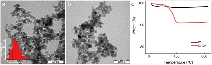
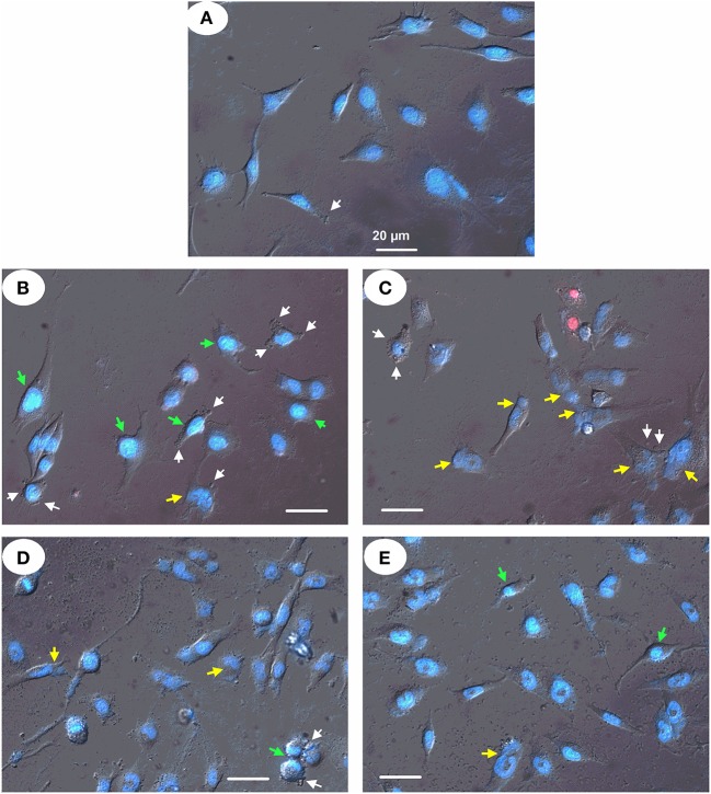
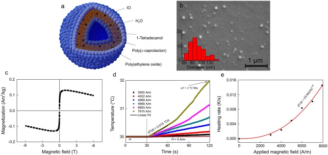
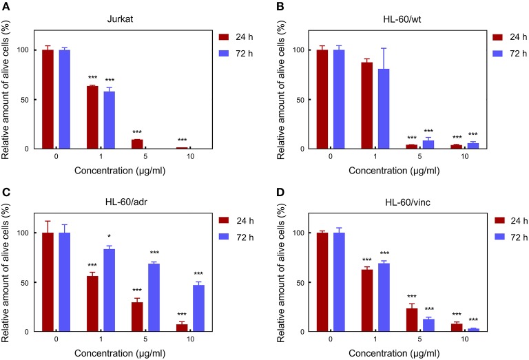
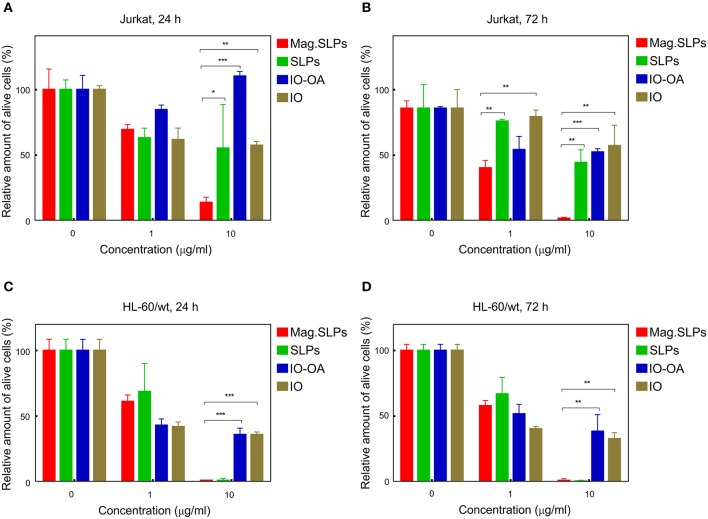
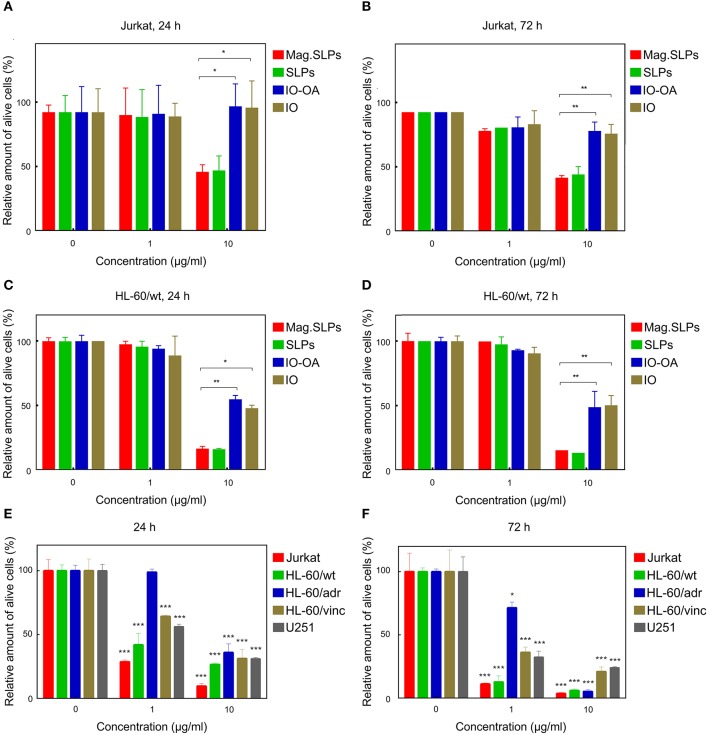
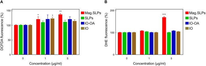
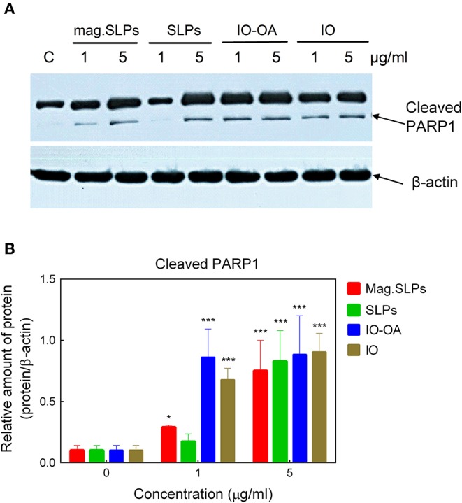
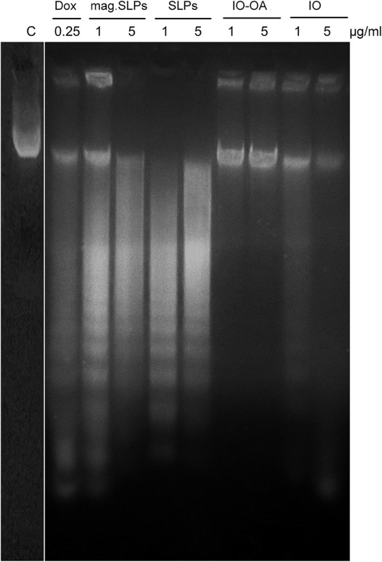
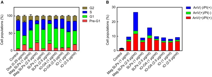
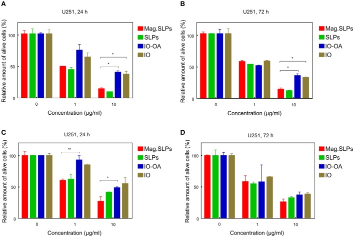
Magnetic and temperature-sensitive solid lipid particles (mag. SLPs) were prepared in the presence of oleic acid-coated iron oxide (IO-OA) nanoparticles with 1-tetradecanol and poly(ethylene oxide)--poly(ε-caprolactone) as lipid and stabilizing surfactant-like agents, respectively. The particles, typically ~850 nm in hydrodynamic size, showed heat dissipation under the applied alternating magnetic field. Cytotoxic activity of the mag.SLPs, non-magnetic SLPs, and iron oxide nanoparticles was compared concerning the mammalian cancer cell lines and their drug-resistant counterparts using trypan blue exclusion test and MTT assay. The mag.SLPs exhibited dose-dependent cytotoxicity against human leukemia cell lines growing in suspension (Jurkat and HL-60/wt), as well as the doxorubicin (Dox)- and vincristine-resistant HL-60 sublines. The mag.SLPs showed higher cytotoxicity toward drug-resistant sublines as compared to Dox. The human glioblastoma cell line U251 growing in a monolayer culture was also sensitive to mag.SLPs cytotoxicity. Staining of U251 cells with the fluorescent dyes Hoechst 33342 and propidium iodide (PI) revealed that mag.SLPs treatment resulted in an increased number of cells with condensed chromatin and/or fragmented nuclei as well as with blebbing of the plasma membranes. While the Hoechst 33342 staining of cell suggested the pro-apoptotic activity of the particles, the PI staining indicated the pro-necrotic changes in the target cells. These conclusions were confirmed by Western blot analysis of apoptosis-related proteins, study of DNA fragmentation (DNA laddering due to the inter-nucleosomal cleavage and DNA comets due to single strand breaks), as well as by FACS analysis of the patterns of cell cycle distribution (pre-G1 phase) and Annexin V/PI staining of the treated Jurkat cells. The induction of apoptosis or necrosis by the particles used to treat Jurkat cells depended on the dose of the particles. Production of the reactive oxygen species (ROS) was proposed as a potential mechanism of mag.SLPs-induced cytotoxicity. Accordingly, hydrogen peroxide and superoxide radical levels in mag.SLPs-treated Jurkat leukemic cells were increased by ~20-40 and ~70%, respectively. In contrast, the non-magnetic SLPs and neat iron oxides did not influence ROS levels significantly. Thus, the developed mag.SLPs can be used for effective killing of human tumor cells, including drug-resistant ones.
在油酸包覆的氧化铁(IO - OA)纳米颗粒存在的情况下,以十四醇和聚(环氧乙烷)-聚(ε - 己内酯)分别作为脂质和稳定的表面活性剂样试剂,制备了磁性和温度敏感的固体脂质颗粒(mag.SLPs)。这些颗粒的流体动力学尺寸通常约为850nm,在施加的交变磁场下显示出热耗散。使用台盼蓝排斥试验和MTT测定法,比较了mag.SLPs、非磁性SLPs和氧化铁纳米颗粒对哺乳动物癌细胞系及其耐药对应物的细胞毒性活性。mag.SLPs对悬浮生长的人白血病细胞系(Jurkat和HL - 60/wt)以及多柔比星(Dox)和长春新碱耐药的HL - 60亚系表现出剂量依赖性细胞毒性。与Dox相比,mag.SLPs对耐药亚系显示出更高的细胞毒性。单层培养的人胶质母细胞瘤细胞系U251也对mag.SLPs细胞毒性敏感。用荧光染料Hoechst 33342和碘化丙啶(PI)对U251细胞进行染色显示,mag.SLPs处理导致染色质浓缩和/或细胞核碎片化以及质膜起泡的细胞数量增加。虽然细胞的Hoechst 33342染色表明颗粒具有促凋亡活性,但PI染色表明靶细胞发生了促坏死变化。通过凋亡相关蛋白的蛋白质印迹分析、DNA片段化研究(由于核小体间切割导致的DNA梯状条带和由于单链断裂导致的DNA彗星)以及对处理后的Jurkat细胞的细胞周期分布模式(G1期前)和膜联蛋白V/PI染色的流式细胞术分析,证实了这些结论。用于处理Jurkat细胞的颗粒诱导凋亡或坏死取决于颗粒的剂量。活性氧(ROS)的产生被认为是mag.SLPs诱导细胞毒性的潜在机制。因此,mag.SLPs处理的Jurkat白血病细胞中过氧化氢和超氧阴离子自由基水平分别增加了约20 - 40%和约70%。相比之下,非磁性SLPs和纯氧化铁对ROS水平没有显著影响。因此,所开发的mag.SLPs可用于有效杀死人肿瘤细胞,包括耐药细胞。