Nie Ji, Ling Wenwu, Yang Qianru, Jin Hongyu, Ou Xuejin, Ma Xuelei
Department of Biotherapy, State Key Laboratory of Biotherapy and Cancer Center, West China Hospital, Sichuan University and Collaborative Innovation Center of Biotherapy, Chengdu, China.
West China School of Medicine, Sichuan University, Chengdu, China.
Front Oncol. 2020 Apr 21;10:473. doi: 10.3389/fonc.2020.00473. eCollection 2020.
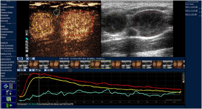
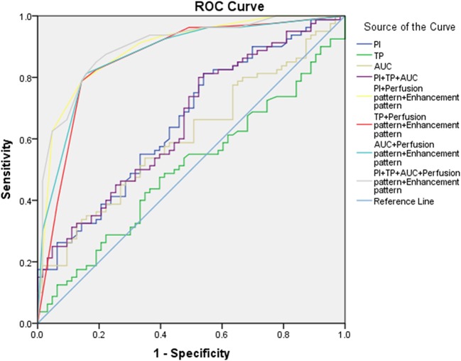
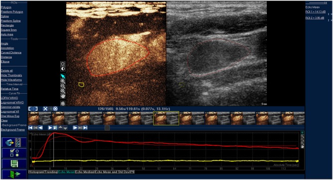
The purpose of this study was to assess the ability of contrast-enhanced ultrasonography (CEUS) in the differential diagnosis of cancerous lymph nodes. Contrast-enhanced ultrasonography was performed in the cervical nodules of included patients, and the diagnoses were confirmed by pathological examination. Contrast-enhanced ultrasonography images and parameters of head and neck lymphomas were compared with those of cancerous lymph nodes. Besides, receiver operating characteristic curve was operated to access the diagnostic value of CEUS. Finally, a total of 63 head and neck lymphomas and 80 cervical cancerous lymph nodes were enrolled in this study. Results showed that the CEUS images of lymphoma were mainly characterized by homogeneous enhancement (71.43%), and approximately half of them were centripetal perfusion (58.73%), whereas most CEUS images of cancerous lymph nodes were inhomogeneous enhancement (82.50%) and centripetal perfusion (92.50%). Quantitative analysis of CEUS parameters indicated that PI (derived peak intensity) and AUC (area under the curve) of lymphomas were both lower than those of cancerous lymph nodes (PI: 8.78 vs. 10.51, AUC: 652.62 vs. 784.09, respectively) ( < 0.05). Receiver operating characteristic analysis showed that the sensitivity of CEUS parameters in the differential diagnosis was significant (80.00%), although the specificity was not high (47.62%). When parameters were combined with the image features, the accuracy of diagnosis was greatly improved (from 0.655 to 0.899). Contrast-enhanced ultrasonography could be a promising tool for the differential diagnosis of head and neck lymphomas and cancerous lymph nodes.
本研究的目的是评估超声造影(CEUS)在癌性淋巴结鉴别诊断中的能力。对纳入患者的颈部结节进行超声造影检查,并通过病理检查确诊。将头颈部淋巴瘤的超声造影图像及参数与癌性淋巴结的进行比较。此外,绘制受试者工作特征曲线以评估CEUS的诊断价值。最终,本研究共纳入63名头颈部淋巴瘤患者和80例颈部癌性淋巴结患者。结果显示,淋巴瘤的CEUS图像主要表现为均匀增强(71.43%),其中约一半为向心性灌注(58.73%),而癌性淋巴结的大多数CEUS图像为不均匀增强(82.50%)和向心性灌注(92.50%)。CEUS参数的定量分析表明,淋巴瘤的PI(峰值强度)和AUC(曲线下面积)均低于癌性淋巴结(PI:8.78对10.51,AUC:652.62对784.09,均P<0.05)。受试者工作特征分析表明,CEUS参数在鉴别诊断中的敏感性显著(80.00%),尽管特异性不高(47.62%)。当参数与图像特征相结合时,诊断准确性大大提高(从0.655提高到0.899)。超声造影可能是头颈部淋巴瘤和癌性淋巴结鉴别诊断的一种有前景的工具。