Department of Biology, University of North Carolina at Chapel Hill, Chapel Hill, NC 27599.
Institute for Systems Genetics, New York University Langone Health, New York, NY 10016.
Mol Biol Cell. 2020 Jul 1;31(14):1498-1511. doi: 10.1091/mbc.E20-03-0210. Epub 2020 May 13.
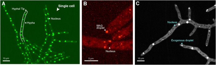
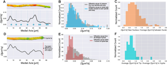
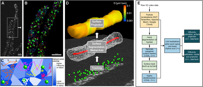
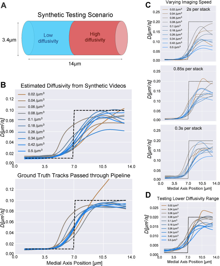
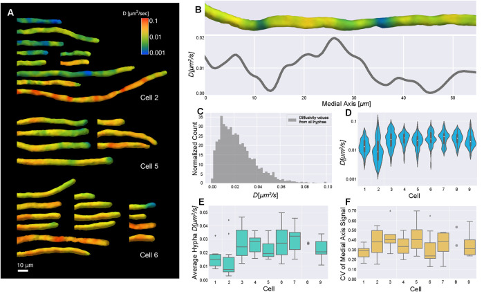
The spatial structure and physical properties of the cytosol are not well understood. Measurements of the material state of the cytosol are challenging due to its spatial and temporal heterogeneity. Recent development of genetically encoded multimeric nanoparticles (GEMs) has opened up study of the cytosol at the length scales of multiprotein complexes (20-60 nm). We developed an image analysis pipeline for 3D imaging of GEMs in the context of large, multinucleate fungi where there is evidence of functional compartmentalization of the cytosol for both the nuclear division cycle and branching. We applied a neural network to track particles in 3D and then created quantitative visualizations of spatially varying diffusivity. Using this pipeline to analyze spatial diffusivity patterns, we found that there is substantial variability in the properties of the cytosol. We detected zones where GEMs display especially low diffusivity at hyphal tips and near some nuclei, showing that the physical state of the cytosol varies spatially within a single cell. Additionally, we observed significant cell-to-cell variability in the average diffusivity of GEMs. Thus, the physical properties of the cytosol vary substantially in time and space and can be a source of heterogeneity within individual cells and across populations.
细胞质的空间结构和物理性质还不太清楚。由于细胞质具有时空异质性,因此对其物质状态进行测量具有挑战性。最近,基因编码的多聚体纳米颗粒(GEMs)的发展为研究多蛋白复合物(20-60nm)长度尺度的细胞质开辟了新途径。我们开发了一种用于在大型多核真菌中进行 GEMs 三维成像的图像分析管道,其中有证据表明细胞质在核分裂周期和分支过程中存在功能分区。我们应用神经网络来追踪三维中的粒子,然后创建空间变化扩散率的定量可视化。使用该管道分析空间扩散率模式,我们发现细胞质的性质存在很大差异。我们检测到在菌丝尖端和靠近某些核附近,GEMs 显示出特别低扩散率的区域,表明细胞质的物理状态在单个细胞内空间上发生变化。此外,我们观察到 GEMs 的平均扩散率在细胞间存在显著差异。因此,细胞质的物理性质在时间和空间上都有很大的变化,并且可能是单个细胞内和细胞群体间异质性的一个来源。