Levada Kateryna, Pshenichnikov Stanislav, Omelyanchik Alexander, Rodionova Valeria, Nikitin Aleksey, Savchenko Alexander, Schetinin Igor, Zhukov Dmitry, Abakumov Maxim, Majouga Alexander, Lunova Mariia, Jirsa Milan, Smolková Barbora, Uzhytchak Mariia, Dejneka Alexandr, Lunov Oleg
Institute of Physics, Mathematics and Information Technology, Immanuel Kant Baltic Federal University, Kaliningrad, Russia.
National University of Science and Technology "MISIS", Moscow, Russia.
Nano Converg. 2020 May 19;7(1):17. doi: 10.1186/s40580-020-00228-5.
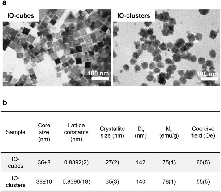
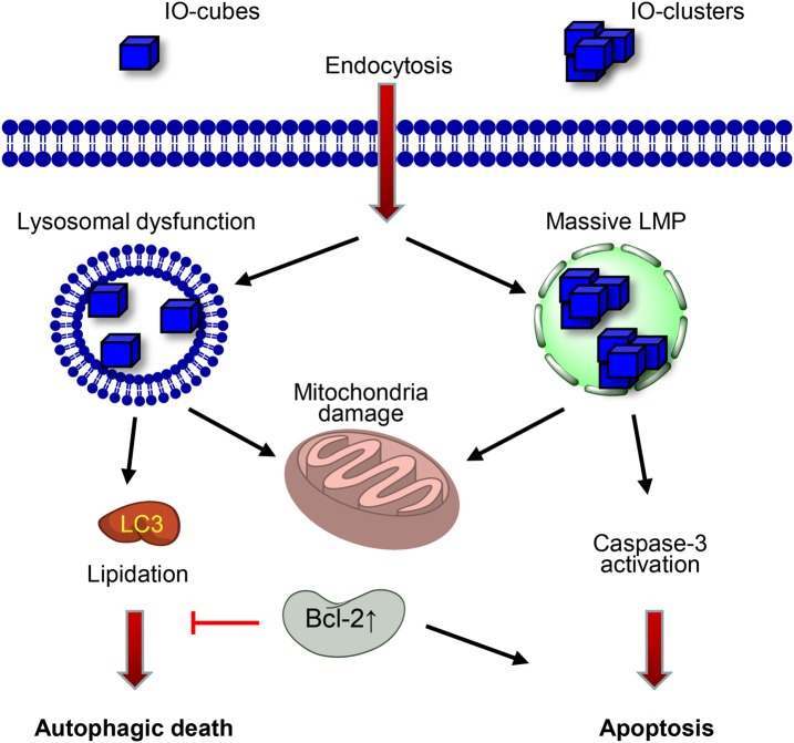
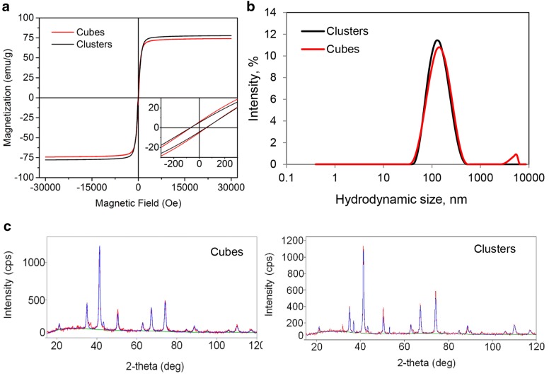
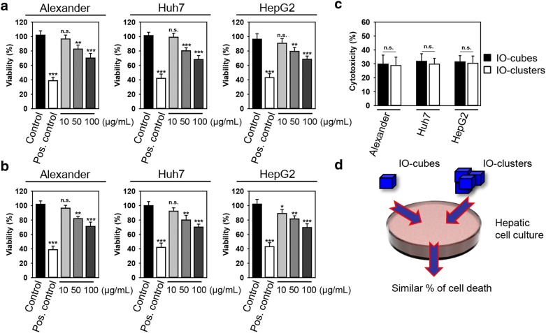
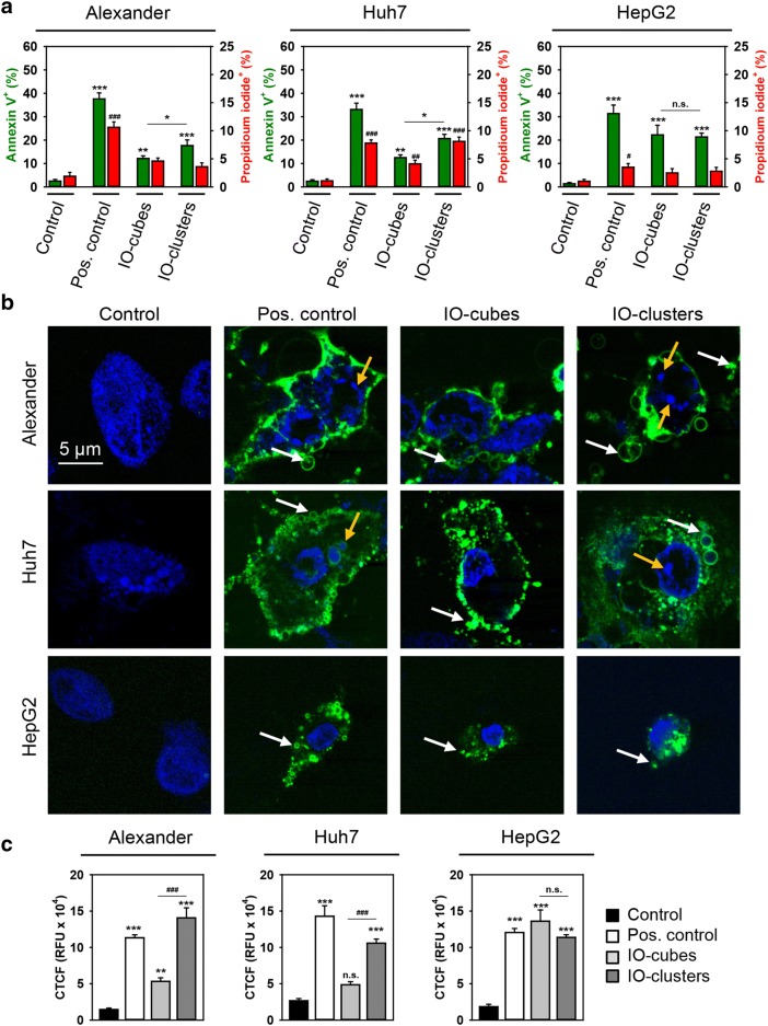
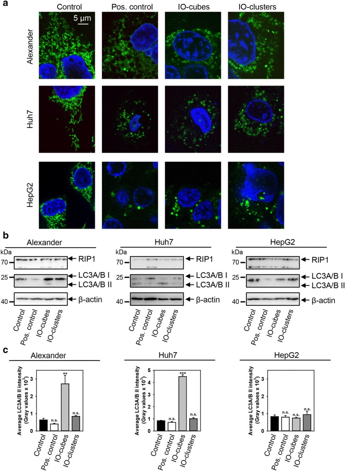
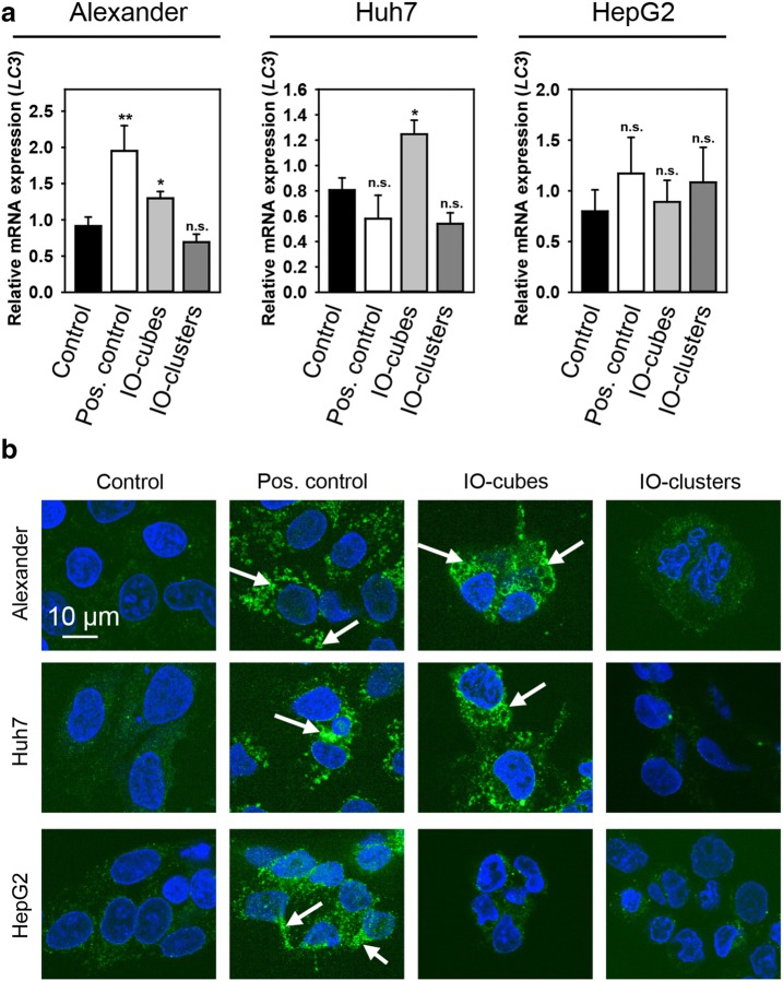
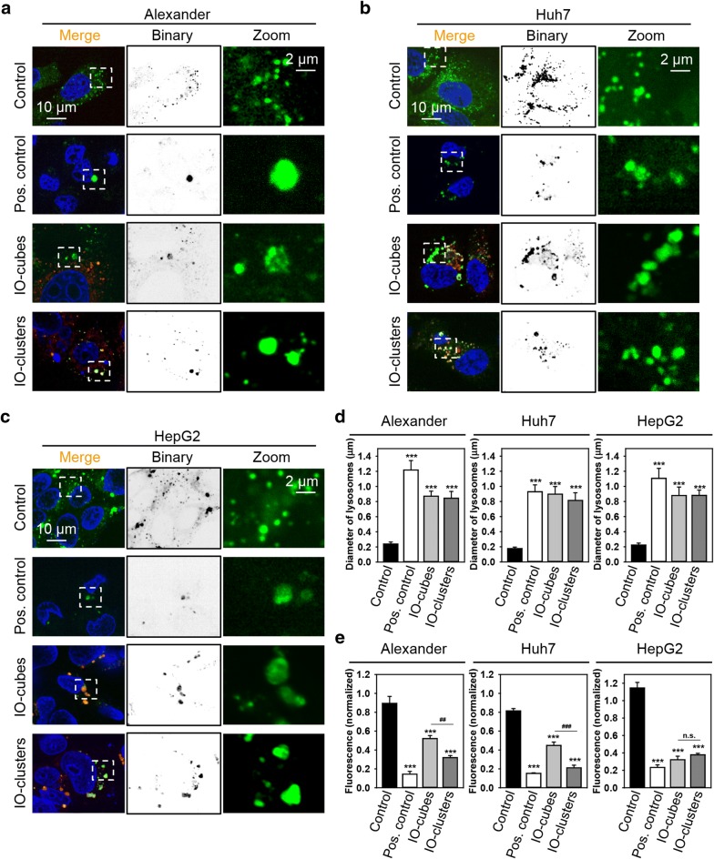
Iron oxide nanoparticles (IONs) are frequently used in various biomedical applications, in particular as magnetic resonance imaging contrast agents in liver imaging. Indeed, number of IONs have been withdrawn due to their poor clinical performance. Yet comprehensive understanding of their interactions with hepatocytes remains relatively limited. Here we investigated how iron oxide nanocubes (IO-cubes) and clusters of nanocubes (IO-clusters) affect distinct human hepatic cell lines. The viability of HepG2, Huh7 and Alexander cells was concentration-dependently decreased after exposure to either IO-cubes or IO-clusters. We found similar cytotoxicity levels in three cell lines triggered by both nanoparticle formulations. Our data indicate that different expression levels of Bcl-2 predispose cell death signaling mediated by nanoparticles. Both nanoparticles induced rather apoptosis than autophagy in HepG2. Contrary, IO-cubes and IO-clusters trigger distinct cell death signaling events in Alexander and Huh7 cells. Our data clarifies the mechanism by which cubic nanoparticles induce autophagic flux and the mechanism of subsequent toxicity. These findings imply that the cytotoxicity of ION-based contrast agents should be carefully considered, particularly in patients with liver diseases.
氧化铁纳米颗粒(IONs)常用于各种生物医学应用中,尤其是作为肝脏成像中的磁共振成像造影剂。事实上,由于其临床性能不佳,已有多种IONs被撤出市场。然而,对于它们与肝细胞相互作用的全面了解仍然相对有限。在此,我们研究了氧化铁纳米立方体(IO-立方体)和纳米立方体簇(IO-簇)如何影响不同的人类肝细胞系。在暴露于IO-立方体或IO-簇后,HepG2、Huh7和Alexander细胞的活力呈浓度依赖性下降。我们发现两种纳米颗粒制剂在三种细胞系中引发的细胞毒性水平相似。我们的数据表明,Bcl-2的不同表达水平易引发纳米颗粒介导的细胞死亡信号。在HepG2细胞中,两种纳米颗粒均诱导细胞凋亡而非自噬。相反,IO-立方体和IO-簇在Alexander细胞和Huh7细胞中引发不同的细胞死亡信号事件。我们的数据阐明了立方纳米颗粒诱导自噬通量的机制以及随后的毒性机制。这些发现意味着,基于ION的造影剂的细胞毒性应予以仔细考虑,尤其是在肝病患者中。