• 文献检索
  • 文档翻译
  • 深度研究
  • 学术资讯
  • Suppr Zotero 插件Zotero 插件
  • 邀请有礼
  • 套餐&价格
  • 历史记录
应用&插件
Suppr Zotero 插件Zotero 插件浏览器插件Mac 客户端Windows 客户端微信小程序
定价
高级版会员购买积分包购买API积分包
服务
文献检索文档翻译深度研究API 文档MCP 服务
关于我们
关于 Suppr公司介绍联系我们用户协议隐私条款
关注我们

Suppr 超能文献

核心技术专利:CN118964589B侵权必究
粤ICP备2023148730 号-1Suppr @ 2026

文献检索

告别复杂PubMed语法,用中文像聊天一样搜索,搜遍4000万医学文献。AI智能推荐,让科研检索更轻松。

立即免费搜索

文件翻译

保留排版,准确专业,支持PDF/Word/PPT等文件格式,支持 12+语言互译。

免费翻译文档

深度研究

AI帮你快速写综述,25分钟生成高质量综述,智能提取关键信息,辅助科研写作。

立即免费体验

同步辐射μCT揭示慢性压迫性胸段脊髓损伤大鼠模型中血管和神经网络的三维微观结构改变

SRμCT Reveals 3D Microstructural Alterations of the Vascular and Neuronal Network in a Rat Model of Chronic Compressive Thoracic Spinal Cord Injury.

作者信息

Jiang Liyuan, Cao Yong, Liu Zhen, Ni Shuangfei, Liu Jun, Ha Yoon, Luo Zixiang, Li Chengjun, Liu Shaohua, Li Jingsong, Yin Xianzhen, Wu Tianding, Lu Hongbin, Hu Jianzhong

机构信息

1Department of Spine Surgery, Xiangya Hospital, Central South University, Changsha, China.

2Key Laboratory of Organ Injury, Aging and Regenerative Medicine of Hunan Province, Changsha, China.

出版信息

Aging Dis. 2020 May 9;11(3):603-617. doi: 10.14336/AD.2019.0529. eCollection 2020 May.

DOI:10.14336/AD.2019.0529
PMID:32489705
原文链接:https://pmc.ncbi.nlm.nih.gov/articles/PMC7220295/
Abstract

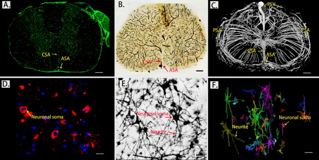
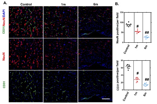
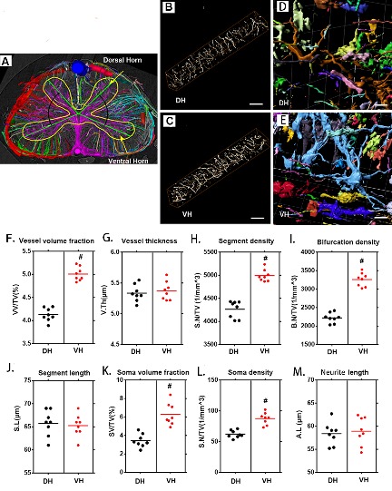
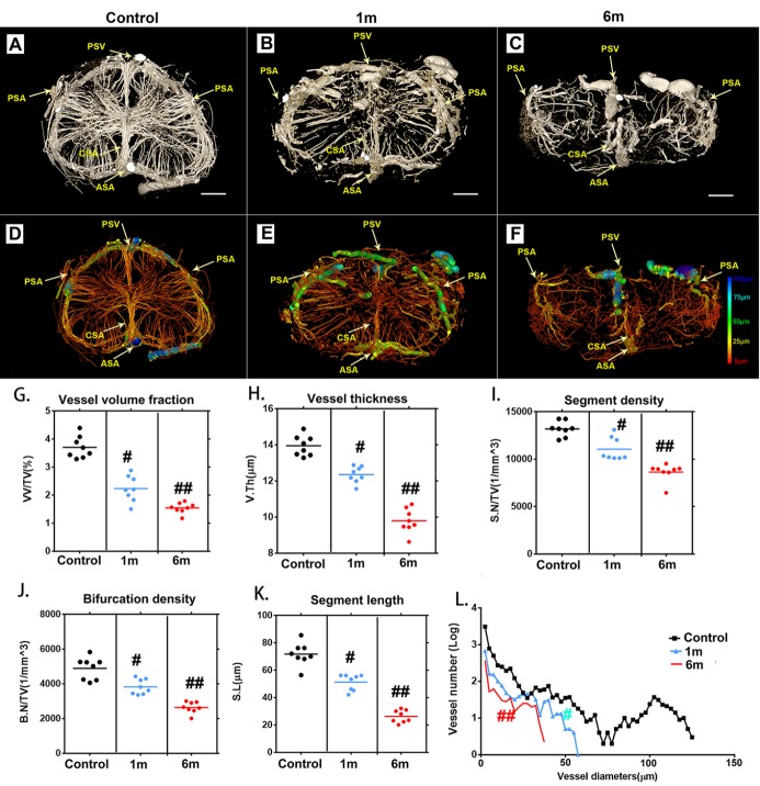
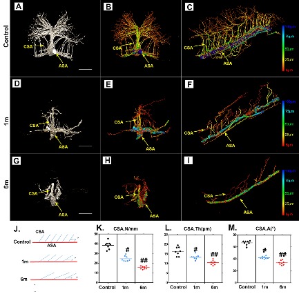
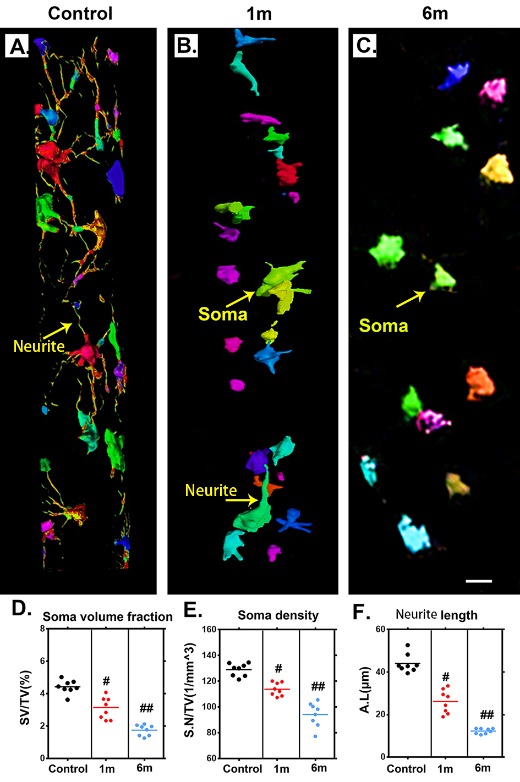
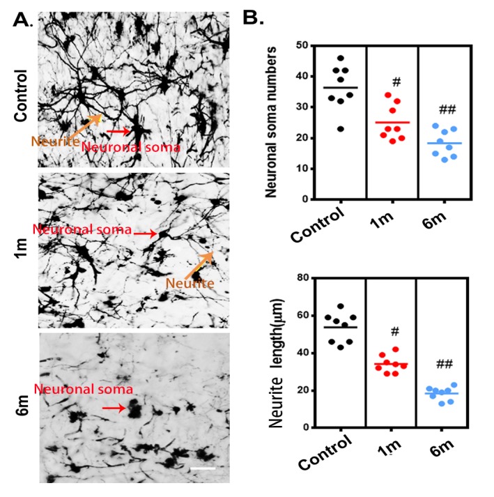
The complex pathology of chronic thoracic spinal cord compression involves vascular and neuroarchitectural repair processes that are still largely unknown. In this study, we used synchrotron radiation microtomography (SRμCT) to quantitatively characterize the 3D temporal-spatial changes in the vascular and neuronal network after chronic thoracic spinal cord compression in order to obtain further insights into the pathogenesis of this disease and to elucidate its underlying mechanisms. Direct 3D characterization of the spinal cord microvasculature and neural microstructure of the thoracic spinal cord was successfully reconstructed. The significant reduction in vasculature and degeneration of neurons in the thoracic spinal cord visualized via SRμCT after chronic compression were consistent with the changes detected by immunofluorescence staining. The 3D morphological measurements revealed significant reductions of neurovascular parameters in the thoracic spinal cord after 1 month of compression and became even worse after 6 months without relief of compression. In addition, the distinct 3D morphological twist and the decrease in branches of the central sulcal artery after chronic compression vividly displayed that these could be the potential triggers leading to blood flow reduction and neural deficits of the thoracic spinal cord. Our findings propose a novel methodology for the 3D analysis of neurovascular repair in chronic spinal cord compression, both qualitatively and quantitatively. The results indicated that compression simultaneously caused vascular dysfunction and neuronal network impairment, which should be acknowledged as concurrent events after chronic thoracic spinal cord injury. Combining neuroprotection with vasoprotection may provide promising therapeutic targets for chronic thoracic spinal cord compression.

摘要

慢性胸段脊髓压迫的复杂病理过程涉及血管和神经结构修复过程,而这些过程在很大程度上仍不为人知。在本研究中,我们使用同步辐射显微断层扫描(SRμCT)对慢性胸段脊髓压迫后血管和神经元网络的三维时空变化进行定量表征,以便进一步深入了解该疾病的发病机制并阐明其潜在机制。成功重建了胸段脊髓微血管和神经微结构的直接三维表征。慢性压迫后通过SRμCT可视化的胸段脊髓血管显著减少和神经元变性与免疫荧光染色检测到的变化一致。三维形态学测量显示,压迫1个月后胸段脊髓的神经血管参数显著降低,在6个月未解除压迫后情况变得更糟。此外,慢性压迫后中央沟动脉明显的三维形态扭曲和分支减少生动地表明,这些可能是导致胸段脊髓血流减少和神经功能缺损的潜在触发因素。我们的研究结果提出了一种用于慢性脊髓压迫中神经血管修复三维分析的新方法,包括定性和定量分析。结果表明,压迫同时导致血管功能障碍和神经元网络损伤,这应被视为慢性胸段脊髓损伤后的并发事件。将神经保护与血管保护相结合可能为慢性胸段脊髓压迫提供有前景的治疗靶点。

https://cdn.ncbi.nlm.nih.gov/pmc/blobs/0a0e/7220295/566f875fe436/ad-11-3-603-g7.jpg
https://cdn.ncbi.nlm.nih.gov/pmc/blobs/0a0e/7220295/9f8fbddcc943/ad-11-3-603-g1.jpg
https://cdn.ncbi.nlm.nih.gov/pmc/blobs/0a0e/7220295/32804506361a/ad-11-3-603-g2.jpg
https://cdn.ncbi.nlm.nih.gov/pmc/blobs/0a0e/7220295/990f00d59aac/ad-11-3-603-g3.jpg
https://cdn.ncbi.nlm.nih.gov/pmc/blobs/0a0e/7220295/2f9e5e57d76c/ad-11-3-603-g4.jpg
https://cdn.ncbi.nlm.nih.gov/pmc/blobs/0a0e/7220295/bdf8ce01fdc7/ad-11-3-603-g5.jpg
https://cdn.ncbi.nlm.nih.gov/pmc/blobs/0a0e/7220295/93c4ecb83b2a/ad-11-3-603-g6.jpg
https://cdn.ncbi.nlm.nih.gov/pmc/blobs/0a0e/7220295/566f875fe436/ad-11-3-603-g7.jpg

相似文献

1
SRμCT Reveals 3D Microstructural Alterations of the Vascular and Neuronal Network in a Rat Model of Chronic Compressive Thoracic Spinal Cord Injury.同步辐射μCT揭示慢性压迫性胸段脊髓损伤大鼠模型中血管和神经网络的三维微观结构改变
Aging Dis. 2020 May 9;11(3):603-617. doi: 10.14336/AD.2019.0529. eCollection 2020 May.
2
Simultaneous 3D Visualization of the Microvascular and Neural Network in Mouse Spinal Cord Using Synchrotron Radiation Micro-Computed Tomography.利用同步辐射微计算机断层扫描技术对小鼠脊髓中的微血管和神经网络进行三维可视化。
Neurosci Bull. 2021 Oct;37(10):1469-1480. doi: 10.1007/s12264-021-00715-7. Epub 2021 Jun 19.
3
3D Digital Anatomic Angioarchitecture of the Rat Spinal Cord: A Synchrotron Radiation Micro-CT Study.大鼠脊髓的3D数字解剖血管构筑:同步辐射微计算机断层扫描研究
Front Neuroanat. 2020 Jul 22;14:41. doi: 10.3389/fnana.2020.00041. eCollection 2020.
4
Three Dimensional Quantification of Microarchitecture and Vessel Regeneration by Synchrotron Radiation Microcomputed Tomography in a Rat Model of Spinal Cord Injury.同步辐射显微计算机断层扫描对大鼠脊髓损伤模型中微结构和血管再生的三维定量分析
J Neurotrauma. 2017 Mar 15;34(6):1187-1199. doi: 10.1089/neu.2016.4697. Epub 2016 Dec 2.
5
A combinatorial method to visualize the neuronal network in the mouse spinal cord: combination of a modified Golgi-Cox method and synchrotron radiation micro-computed tomography.一种可视化小鼠脊髓神经元网络的组合方法:改良的高尔基-考克斯法与同步辐射微计算机断层扫描的结合。
Histochem Cell Biol. 2021 Apr;155(4):477-489. doi: 10.1007/s00418-020-01949-8. Epub 2021 Jan 4.
6
Three-dimensional imaging of microvasculature in the rat spinal cord following injury.大鼠脊髓损伤后微血管系统的三维成像
Sci Rep. 2015 Jul 29;5:12643. doi: 10.1038/srep12643.
7
Non-destructive 3D Microtomography of Cerebral Angioarchitecture Changes Following Ischemic Stroke in Rats Using Synchrotron Radiation.使用同步辐射对大鼠缺血性中风后脑血管结构变化进行无损三维显微断层扫描
Front Neuroanat. 2019 Jan 31;13:5. doi: 10.3389/fnana.2019.00005. eCollection 2019.
8
3D visualization and morphometric analysis of spinal motion segments and vascular networks: A synchrotron radiation-based micro-CT study in mice.基于同步辐射的 micro-CT 研究在小鼠中对脊柱运动节段和血管网络的 3D 可视化和形态计量分析。
J Anat. 2022 Feb;240(2):268-278. doi: 10.1111/joa.13556. Epub 2021 Oct 7.
9
Changes in axonal physiology and morphology after chronic compressive injury of the rat thoracic spinal cord.大鼠胸段脊髓慢性压迫性损伤后轴突生理和形态的变化
Neuroscience. 2001;104(1):235-51. doi: 10.1016/s0306-4522(01)00009-4.
10
Neuronal loss and expression of neurotrophic factors in a model of rat chronic compressive spinal cord injury.大鼠慢性压迫性脊髓损伤模型中的神经元丢失及神经营养因子表达
Spine (Phila Pa 1976). 2006 Aug 15;31(18):2059-66. doi: 10.1097/01.brs.0000231893.21964.f2.

引用本文的文献

1
Loading tea polyphenols enhances the repair of human umbilical cord mesenchymal stem cell sheet after spinal cord injury.负载茶多酚可增强脊髓损伤后人脐带间充质干细胞片的修复能力。
Stem Cell Res Ther. 2025 May 28;16(1):264. doi: 10.1186/s13287-025-04376-5.
2
Neurovascular Unit Compensation from Adjacent Level May Contribute to Spontaneous Functional Recovery in Experimental Cervical Spondylotic Myelopathy.相邻节段的神经血管单元代偿可能有助于实验性脊髓型颈椎病的自发功能恢复。
Int J Mol Sci. 2023 Feb 8;24(4):3408. doi: 10.3390/ijms24043408.
3
Limb Remote Ischemic Conditioning Promotes Neurogenesis after Cerebral Ischemia by Modulating miR-449b/Notch1 Pathway in Mice.

本文引用的文献

1
Correction: Effects of Low-Intensity Pulsed Ultrasound on New Trabecular Bone during Bone-Tendon Junction Healing in a Rabbit Model: A Synchrotron Radiation Micro-CT Study.更正:低强度脉冲超声对兔模型骨-肌腱结合部愈合过程中新小梁骨的影响:同步辐射显微CT研究
PLoS One. 2019 Apr 1;14(4):e0214974. doi: 10.1371/journal.pone.0214974. eCollection 2019.
2
Non-destructive 3D Microtomography of Cerebral Angioarchitecture Changes Following Ischemic Stroke in Rats Using Synchrotron Radiation.使用同步辐射对大鼠缺血性中风后脑血管结构变化进行无损三维显微断层扫描
Front Neuroanat. 2019 Jan 31;13:5. doi: 10.3389/fnana.2019.00005. eCollection 2019.
3
肢体远程缺血预处理通过调节 miR-449b/Notch1 通路促进脑缺血后神经发生。
Biomolecules. 2022 Aug 18;12(8):1137. doi: 10.3390/biom12081137.
4
Ultrafast Doppler imaging and ultrasound localization microscopy reveal the complexity of vascular rearrangement in chronic spinal lesion.超快速多普勒成像和超声定位显微镜揭示慢性脊髓病变中血管重排的复杂性。
Sci Rep. 2022 Apr 21;12(1):6574. doi: 10.1038/s41598-022-10250-8.
5
Bone Marrow Mesenchymal Stem Cell-Derived Exosome-Educated Macrophages Promote Functional Healing After Spinal Cord Injury.骨髓间充质干细胞来源的外泌体诱导巨噬细胞促进脊髓损伤后的功能修复。
Front Cell Neurosci. 2021 Sep 28;15:725573. doi: 10.3389/fncel.2021.725573. eCollection 2021.
6
Microglia-Derived Exosomes Improve Spinal Cord Functional Recovery after Injury via Inhibiting Oxidative Stress and Promoting the Survival and Function of Endothelia Cells.小胶质细胞衍生的外泌体通过抑制氧化应激和促进内皮细胞的存活和功能来改善损伤后脊髓的功能恢复。
Oxid Med Cell Longev. 2021 Aug 25;2021:1695087. doi: 10.1155/2021/1695087. eCollection 2021.
7
Limb Remote Ischemic Conditioning Ameliorates Cognitive Impairment in Rats with Chronic Cerebral Hypoperfusion by Regulating Glucose Transport.肢体远程缺血预处理通过调节葡萄糖转运改善慢性脑灌注不足大鼠的认知障碍。
Aging Dis. 2021 Aug 1;12(5):1197-1210. doi: 10.14336/AD.2020.1125. eCollection 2021 Aug.
The role of brain vasculature in neurodegenerative disorders.
脑血管在神经退行性疾病中的作用。
Nat Neurosci. 2018 Oct;21(10):1318-1331. doi: 10.1038/s41593-018-0234-x. Epub 2018 Sep 24.
4
Apoε4 disrupts neurovascular regulation and undermines white matter integrity and cognitive function.载脂蛋白 E4 破坏神经血管调节,破坏大脑白质完整性和认知功能。
Nat Commun. 2018 Sep 19;9(1):3816. doi: 10.1038/s41467-018-06301-2.
5
Synchrotron Radiation Imaging Reveals the Role of Estrogen in Promoting Angiogenesis After Acute Spinal Cord Injury in Rats.同步辐射成像揭示了雌激素在促进大鼠急性脊髓损伤后血管生成中的作用。
Spine (Phila Pa 1976). 2018 Sep 15;43(18):1241-1249. doi: 10.1097/BRS.0000000000002629.
6
Ultrastructural Features of Neurovascular Units in a Rat Model of Chronic Compressive Spinal Cord Injury.慢性压迫性脊髓损伤大鼠模型中神经血管单元的超微结构特征
Front Neuroanat. 2018 Jan 10;11:136. doi: 10.3389/fnana.2017.00136. eCollection 2017.
7
Three-dimensional visualization and pathologic characteristics of cartilage and subchondral bone changes in the lumbar facet joint of an ovariectomized mouse model.去卵巢小鼠腰椎小关节软骨及软骨下骨改变的三维可视化及病理学特征。
Spine J. 2018 Apr;18(4):663-673. doi: 10.1016/j.spinee.2017.11.009. Epub 2017 Nov 15.
8
Histological, immunohistochemical and ultrastructural study of secondary compressed spinal cord injury in a rat model.大鼠模型中继发性压迫性脊髓损伤的组织学、免疫组织化学及超微结构研究
Folia Histochem Cytobiol. 2017;55(1):11-20. doi: 10.5603/FHC.a2017.0001.
9
3D characterization of morphological changes in the intervertebral disc and endplate during aging: A propagation phase contrast synchrotron micro-tomography study.3D 特征化研究:在衰老过程中椎间盘和终板形态变化的传播:相位对比同步辐射微断层扫描研究。
Sci Rep. 2017 Mar 7;7:43094. doi: 10.1038/srep43094.
10
Comparison of Synchrotron Radiation-based Propagation Phase Contrast Imaging and Conventional Micro-computed Tomography for Assessing Intervertebral Discs and Endplates in a Murine Model.基于同步辐射的传播相位对比成像与传统微计算机断层扫描在评估小鼠模型椎间盘和终板中的比较
Spine (Phila Pa 1976). 2017 Aug 1;42(15):E883-E889. doi: 10.1097/BRS.0000000000002110.