Kim Soyoung, Greene Deanna J, D'Andrea Carolina Badke, Bihun Emily C, Koller Jonathan M, O'Reilly Bridget, Schlaggar Bradley L, Black Kevin J
Department of Psychiatry, Washington University School of Medicine, St. Louis, MO 63110, USA.
Department of Radiology, Washington University School of Medicine, St. Louis, MO 63110, USA.
J Clin Med. 2020 Jun 3;9(6):1715. doi: 10.3390/jcm9061715.
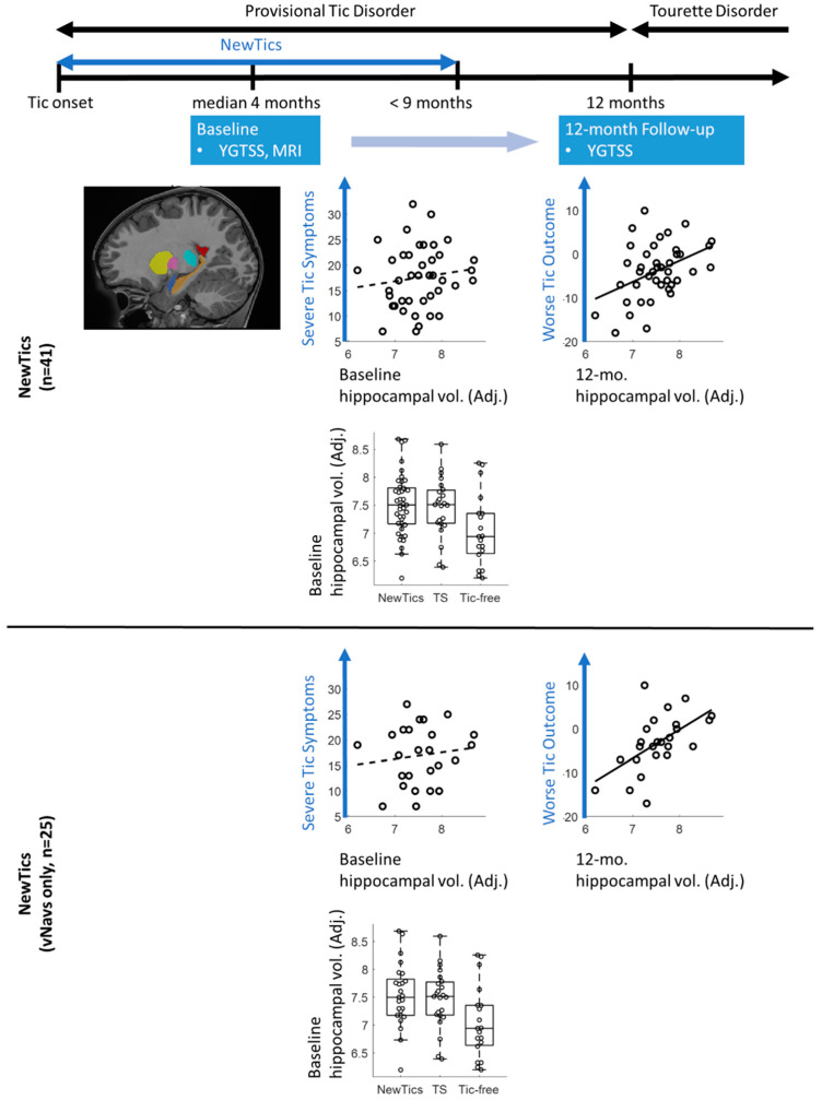
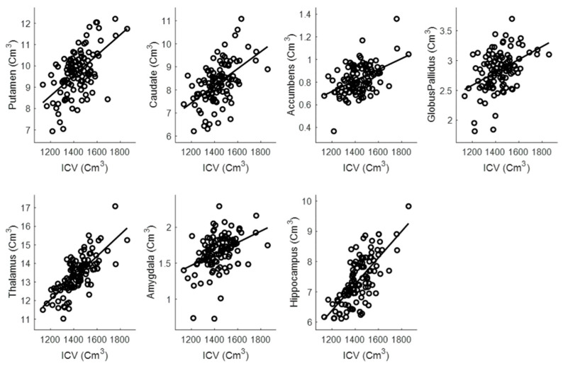
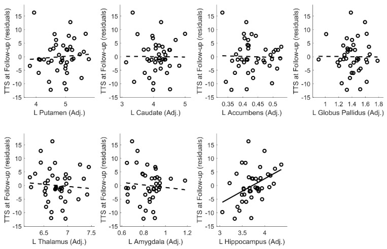
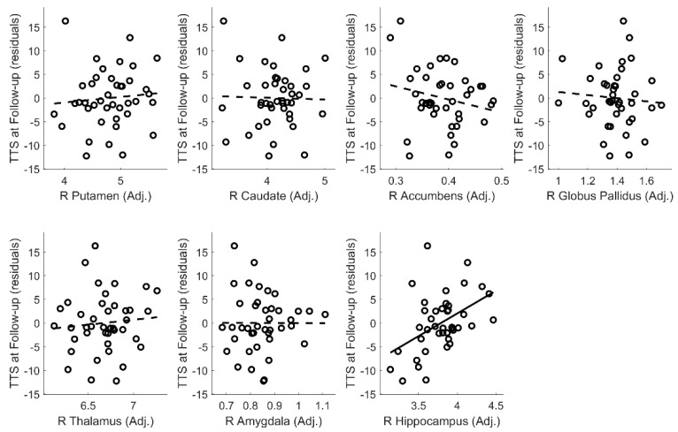
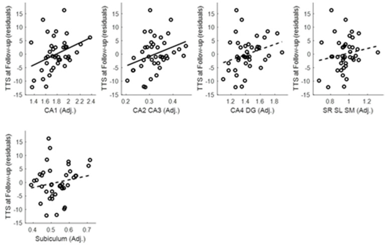
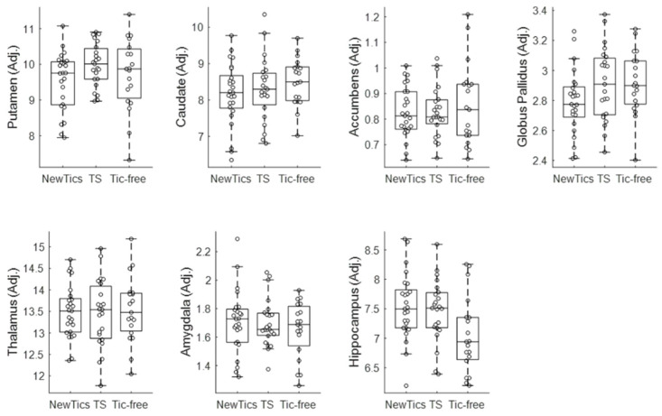
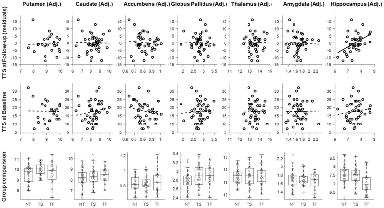
Previous studies have investigated differences in the volumes of subcortical structures (e.g., caudate nucleus, putamen, thalamus, amygdala, and hippocampus) between individuals with and without Tourette syndrome (TS), as well as the relationships between these volumes and tic symptom severity. These volumes may also predict clinical outcome in Provisional Tic Disorder (PTD), but that hypothesis has never been tested. This study aimed to examine whether the volumes of subcortical structures measured shortly after tic onset can predict tic symptom severity at one-year post-tic onset, when TS can first be diagnosed. We obtained T1-weighted structural MRI scans from 41 children with PTD (25 with prospective motion correction (vNavs)) whose tics had begun less than 9 months (mean 4.04 months) prior to the first study visit (baseline). We re-examined them at the 12-month anniversary of their first tic (follow-up), assessing tic severity using the Yale Global Tic Severity Scale. We quantified the volumes of subcortical structures using volBrain software. Baseline hippocampal volume was correlated with tic severity at the 12-month follow-up, with a larger hippocampus at baseline predicting worse tic severity at follow-up. The volumes of other subcortical structures did not significantly predict tic severity at follow-up. Hippocampal volume may be an important marker in predicting prognosis in Provisional Tic Disorder.
以往的研究调查了患有和未患有妥瑞氏症(TS)的个体之间皮质下结构(如尾状核、壳核、丘脑、杏仁核和海马体)体积的差异,以及这些体积与抽动症状严重程度之间的关系。这些体积也可能预测暂时性抽动障碍(PTD)的临床结局,但这一假设从未得到验证。本研究旨在探讨在抽动发作后不久测量的皮质下结构体积是否能够预测抽动发作一年后(此时TS首次可被诊断)的抽动症状严重程度。我们从41名患有PTD的儿童(其中25名采用前瞻性运动校正(vNavs))获取了T1加权结构MRI扫描图像,这些儿童的抽动在首次研究访视(基线)前不到9个月(平均4.04个月)开始。我们在他们首次抽动的12个月纪念日(随访)对他们进行了重新检查,使用耶鲁全球抽动严重程度量表评估抽动严重程度。我们使用volBrain软件对皮质下结构的体积进行了量化。基线时海马体体积与12个月随访时的抽动严重程度相关,基线时较大的海马体预示着随访时更严重的抽动症状。其他皮质下结构的体积在随访时并未显著预测抽动严重程度。海马体体积可能是预测暂时性抽动障碍预后的一个重要指标。