Department of Anesthesiology, Beijing Anzhen Hospital, Capital Medical University-Beijing Institute of Heart Lung and Blood Vessel Diseases, Beijing, People's Republic of China.
Drug Des Devel Ther. 2020 May 25;14:2047-2060. doi: 10.2147/DDDT.S248628. eCollection 2020.
Autophagy caused by ischemia/reperfusion (I/R) increases the extent of cardiomyocyte damage. Melatonin (Mel) diminishes cardiac injury through regulating autophagy and mitochondrial dynamics. However, illustrating the specific role of mitophagy in the cardioprotective effects of melatonin remains a challenge. The aim of our research was to investigate the impact and underlying mechanisms of melatonin in connection with mitophagy during anoxia/reoxygenation (A/R) injury in H9c2 cells.
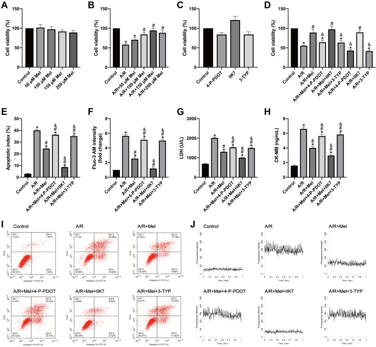
H9c2 cells were pretreated with melatonin with or without the melatonin membrane receptor 2 (MT2) antagonist 4-P-PDOT, the MT2 agonist IIK7 and the sirtuin 3 (SIRT3) inhibitor 3-TYP for 4 hours and then subjected to A/R injury. Cell viability, cellular apoptosis, necrosis levels and oxidative markers were assessed. The expression of SIRT3 and forkhead box O3a (FoxO3a), mitochondrial function and the levels of mitophagy-related proteins were also evaluated.
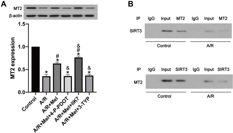
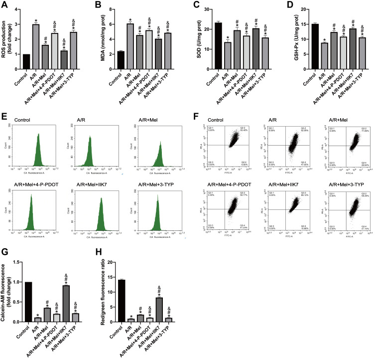
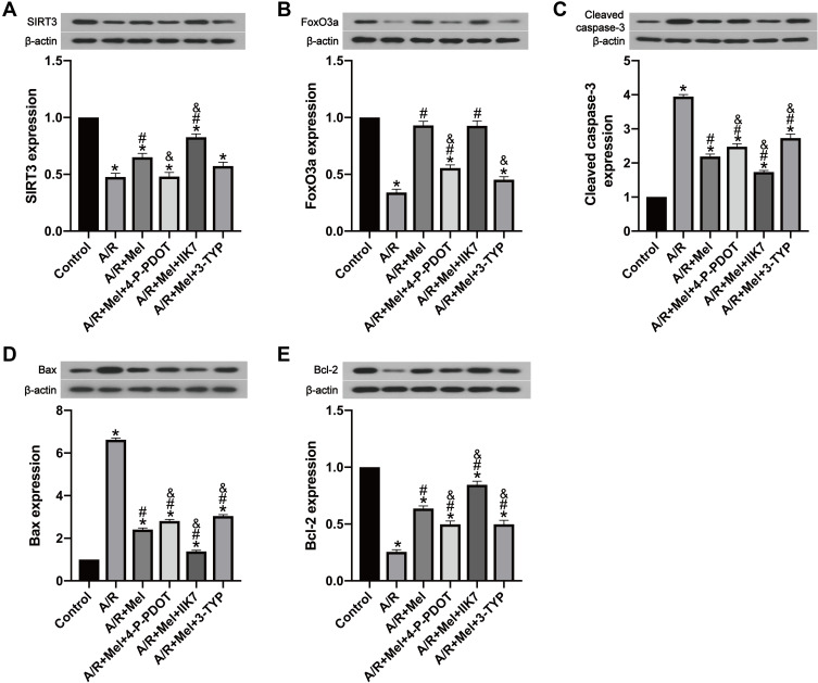
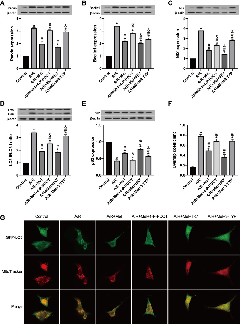
A/R injury provoked enhanced mitophagy in H9c2 myocytes. In addition, increased mitophagy was correlated with decreased cellular viability, increased oxidative stress and mitochondrial dysfunction in H9c2 cells. However, melatonin pretreatment notably increased cell survival and decreased cell apoptosis and oxidative response after A/R injury, accompanied by restored mitochondrial function. The inhibition of excessive mitophagy is involved in the cardioprotective effects of melatonin, as shown by the decreased expression of the mitophagy-related molecules Parkin, Beclin1, and BCL2-interacting protein 3-like (BNIP3L, best known as NIX) and decreased light chain 3 II/light chain 3 I (LC3 II/LC3 I) ratio and upregulation of p62 expression. Moreover, the decreased expression of SIRT3 and FoxO3a in A/R-injured H9c2 cells was abrogated by melatonin, but these beneficial effects were attenuated by the MT2 antagonist 4-P-PDOT or the SIRT3 inhibitor 3-TYP and enhanced by the MT2 agonist IIK7.
These results indicate that melatonin protects H9c2 cells during A/R injury through suppressing excessive mitophagy by activating the MT2/SIRT3/FoxO3a pathway. Melatonin may be a useful candidate for alleviating myocardial ischemia/reperfusion (MI/R) injury in the future, and the MT2 receptor might become a therapeutic target.
缺血/再灌注(I/R)引起的自噬会增加心肌细胞损伤的程度。褪黑素(Mel)通过调节自噬和线粒体动力学来减轻心脏损伤。然而,阐明褪黑素在自噬中的特定作用对于阐明其心脏保护作用仍然是一个挑战。本研究的目的是探讨褪黑素在 H9c2 细胞缺氧/复氧(A/R)损伤过程中与线粒体自噬相关的作用及潜在机制。
H9c2 细胞用褪黑素预处理,或用褪黑素膜受体 2(MT2)拮抗剂 4-P-PDOT、MT2 激动剂 IIK7 和 SIRT3 抑制剂 3-TYP 预处理 4 小时,然后进行 A/R 损伤。检测细胞活力、细胞凋亡、坏死水平和氧化标记物。还评估了 SIRT3 和叉头框 O3a(FoxO3a)、线粒体功能和自噬相关蛋白的水平。
A/R 损伤引起 H9c2 心肌细胞中自噬增加。此外,在 H9c2 细胞中,增加的自噬与细胞活力降低、氧化应激增加和线粒体功能障碍相关。然而,褪黑素预处理显著增加了 A/R 损伤后细胞的存活率,降低了细胞凋亡和氧化反应,同时恢复了线粒体功能。抑制过度的自噬参与了褪黑素的心脏保护作用,表现为自噬相关分子 Parkin、Beclin1 和 BCL2 相互作用蛋白 3 样(BNIP3L,又称 NIX)以及 LC3 II/LC3 I 比值降低和 p62 表达增加的减少。此外,A/R 损伤的 H9c2 细胞中 SIRT3 和 FoxO3a 的表达减少被褪黑素所阻断,但这些有益作用被 MT2 拮抗剂 4-P-PDOT 或 SIRT3 抑制剂 3-TYP 减弱,被 MT2 激动剂 IIK7 增强。
这些结果表明,褪黑素通过激活 MT2/SIRT3/FoxO3a 通路抑制过度自噬来保护 H9c2 细胞在 A/R 损伤期间的活力。褪黑素可能是未来缓解心肌缺血/再灌注(MI/R)损伤的一种有用候选药物,而 MT2 受体可能成为一个治疗靶点。