• 文献检索
  • 文档翻译
  • 深度研究
  • 学术资讯
  • Suppr Zotero 插件Zotero 插件
  • 邀请有礼
  • 套餐&价格
  • 历史记录
应用&插件
Suppr Zotero 插件Zotero 插件浏览器插件Mac 客户端Windows 客户端微信小程序
定价
高级版会员购买积分包购买API积分包
服务
文献检索文档翻译深度研究API 文档MCP 服务
关于我们
关于 Suppr公司介绍联系我们用户协议隐私条款
关注我们

Suppr 超能文献

核心技术专利:CN118964589B侵权必究
粤ICP备2023148730 号-1Suppr @ 2026

文献检索

告别复杂PubMed语法,用中文像聊天一样搜索,搜遍4000万医学文献。AI智能推荐,让科研检索更轻松。

立即免费搜索

文件翻译

保留排版,准确专业,支持PDF/Word/PPT等文件格式,支持 12+语言互译。

免费翻译文档

深度研究

AI帮你快速写综述,25分钟生成高质量综述,智能提取关键信息,辅助科研写作。

立即免费体验

3D 相关低温冷冻结构照明显微镜和软 X 射线显微镜阐明了呼肠孤病毒的细胞内释放途径。

3D Correlative Cryo-Structured Illumination Fluorescence and Soft X-ray Microscopy Elucidates Reovirus Intracellular Release Pathway.

机构信息

Diamond Light Source, Harwell Science and Innovation Campus, Didcot OX11 0DE, UK.

Department of Infectious Diseases, Molecular Virology, Heidelberg University Hospital, 69120 Heidelberg, Germany.

出版信息

Cell. 2020 Jul 23;182(2):515-530.e17. doi: 10.1016/j.cell.2020.05.051. Epub 2020 Jun 30.

DOI:10.1016/j.cell.2020.05.051
PMID:32610083
原文链接:https://pmc.ncbi.nlm.nih.gov/articles/PMC7391008/
Abstract

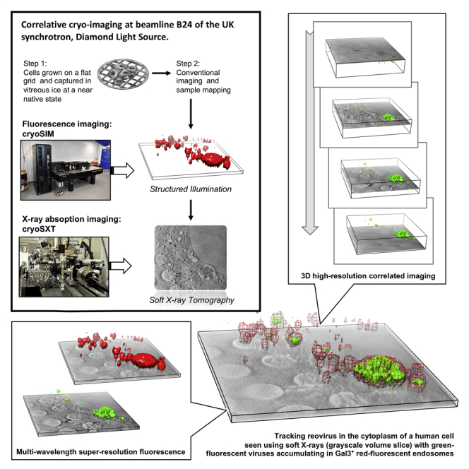
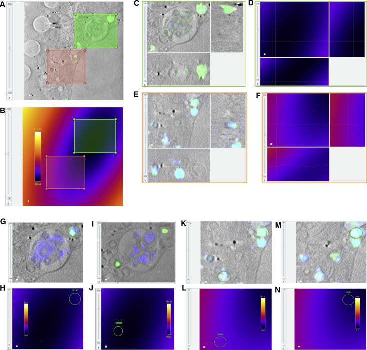
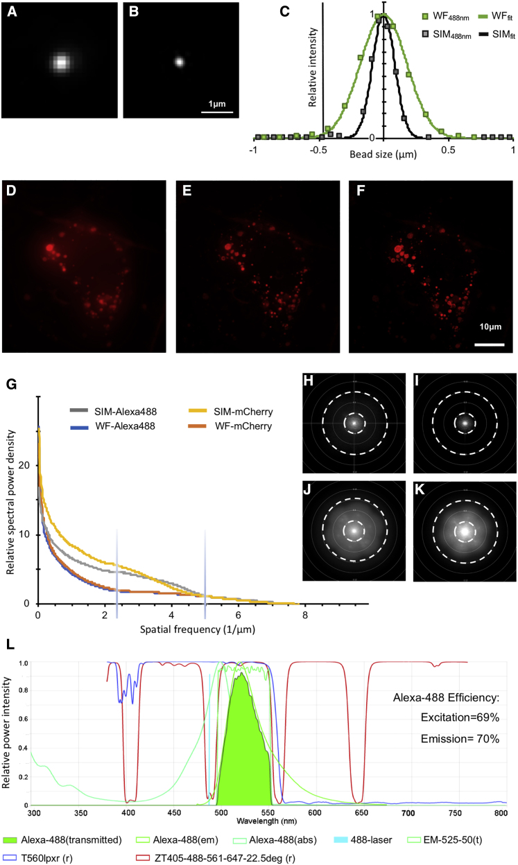
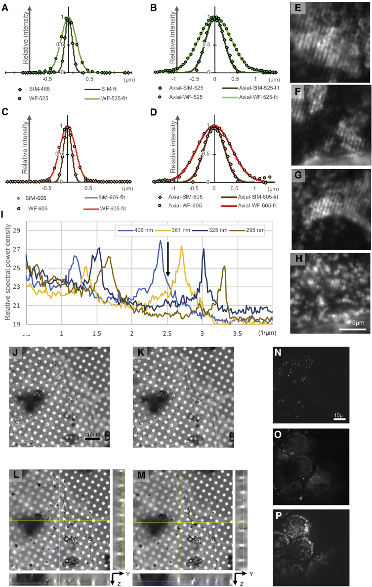
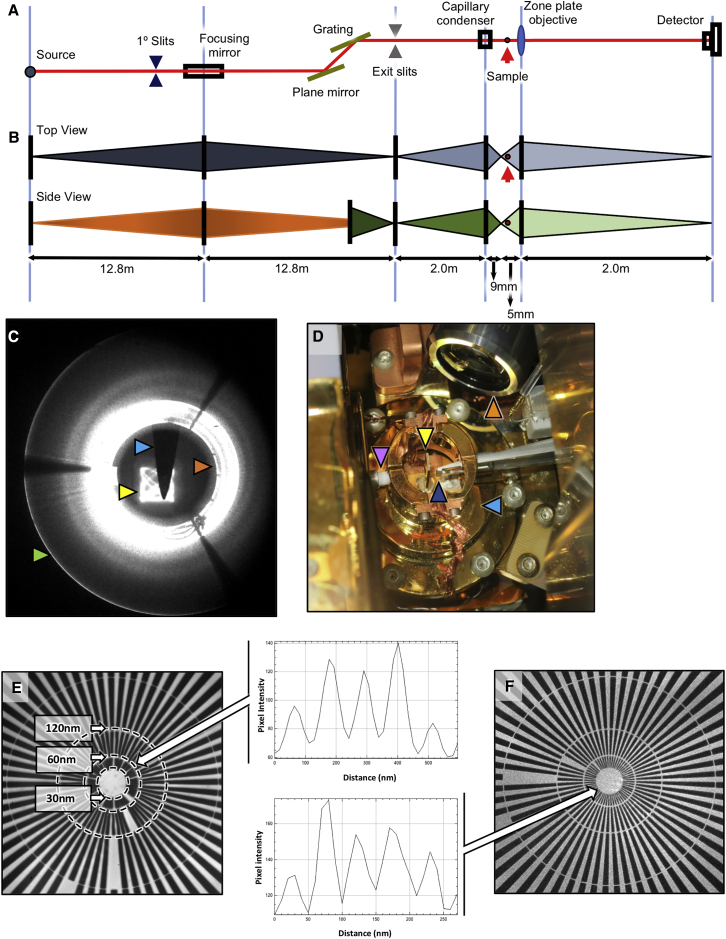
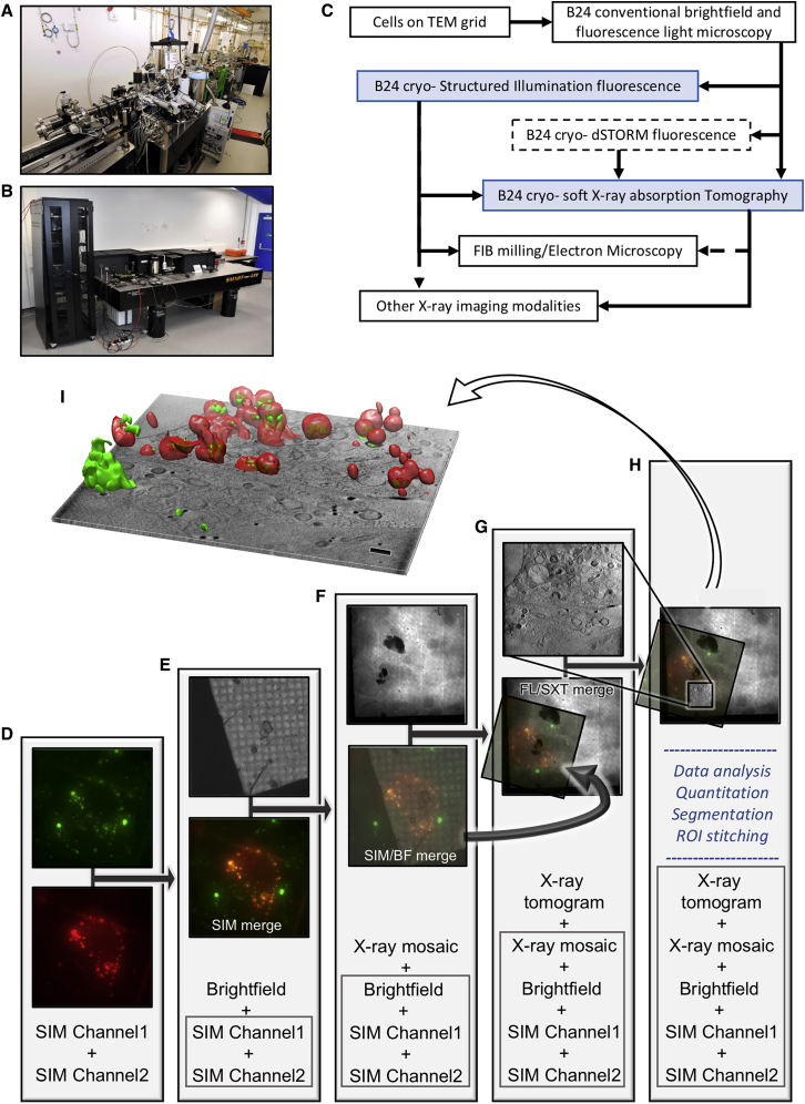
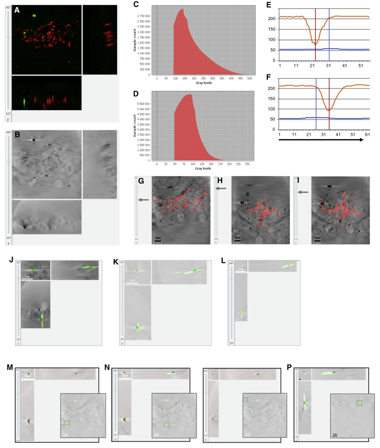
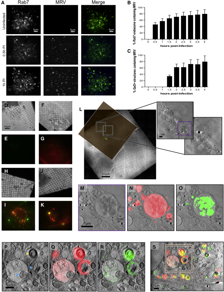
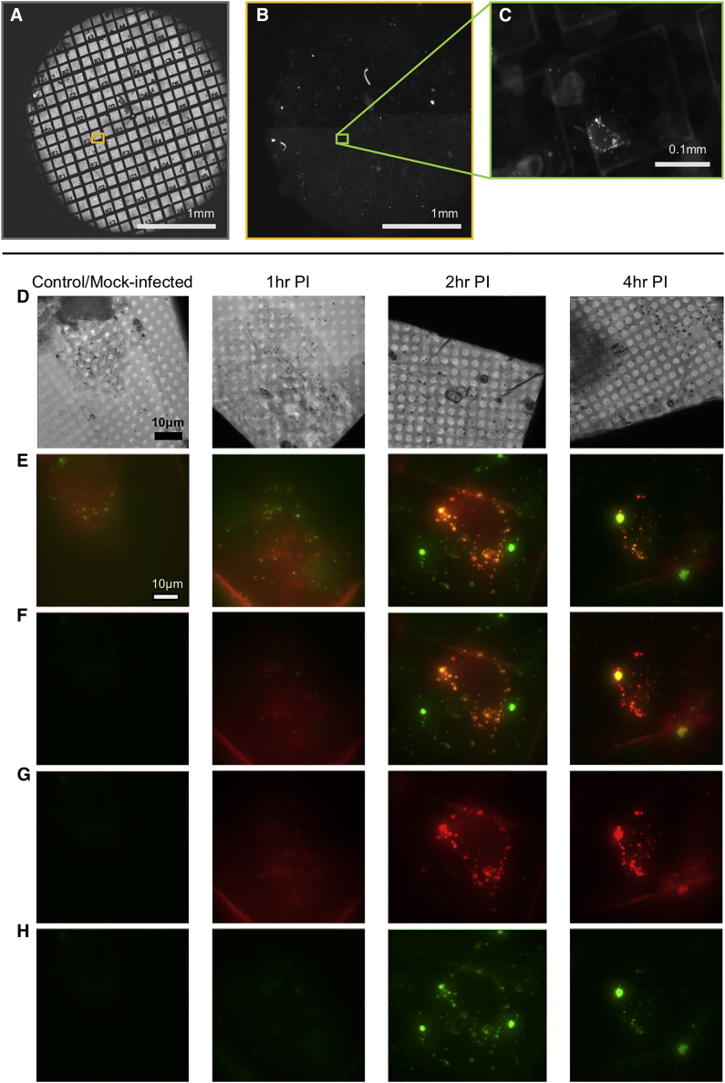
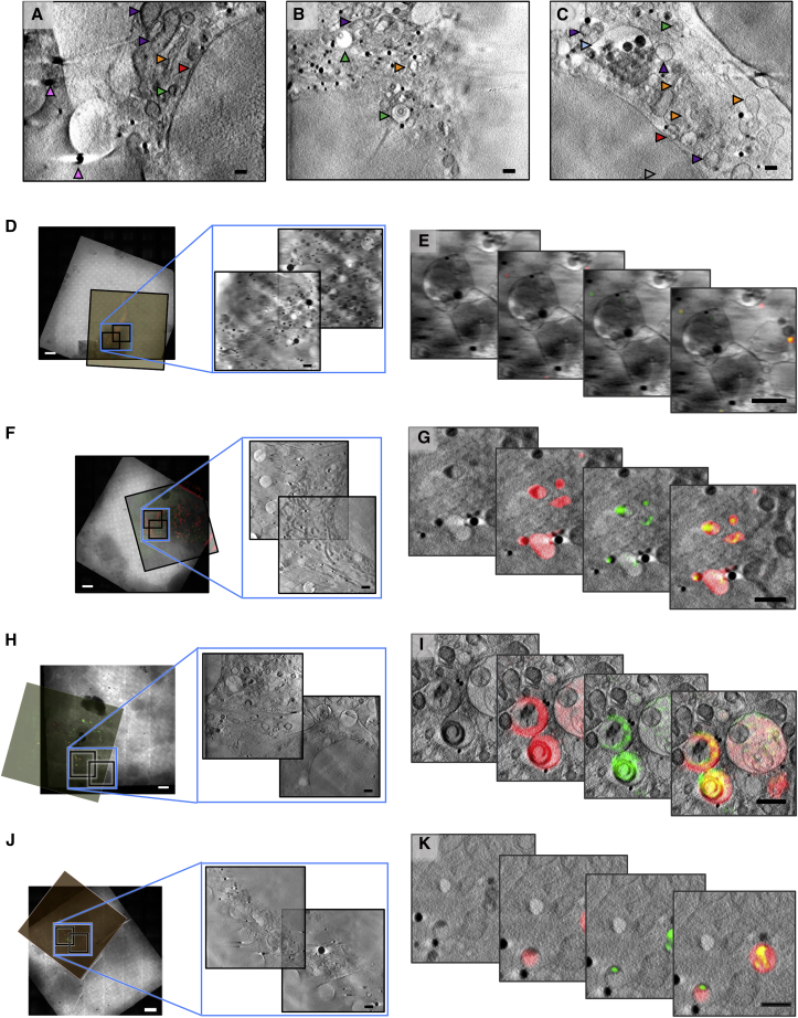
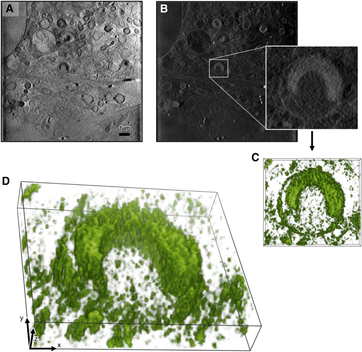
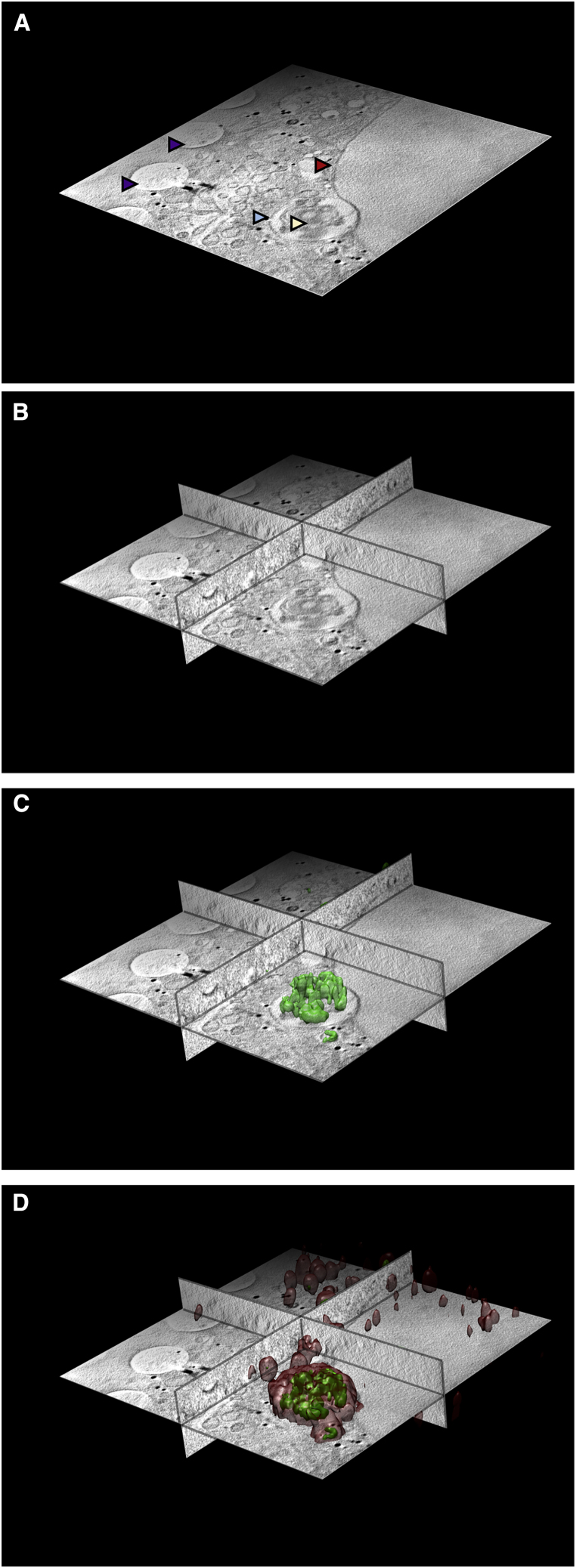
Imaging of biological matter across resolution scales entails the challenge of preserving the direct and unambiguous correlation of subject features from the macroscopic to the microscopic level. Here, we present a correlative imaging platform developed specifically for imaging cells in 3D under cryogenic conditions by using X-rays and visible light. Rapid cryo-preservation of biological specimens is the current gold standard in sample preparation for ultrastructural analysis in X-ray imaging. However, cryogenic fluorescence localization methods are, in their majority, diffraction-limited and fail to deliver matching resolution. We addressed this technological gap by developing an integrated, user-friendly platform for 3D correlative imaging of cells in vitreous ice by using super-resolution structured illumination microscopy in conjunction with soft X-ray tomography. The power of this approach is demonstrated by studying the process of reovirus release from intracellular vesicles during the early stages of infection and identifying intracellular virus-induced structures.

摘要

跨分辨率尺度的生物物质成像是一项具有挑战性的工作,需要确保从宏观到微观层面的主体特征具有直接且明确的相关性。在这里,我们展示了一种专门用于在低温条件下通过 X 射线和可见光对细胞进行三维成像的相关成像平台。快速冷冻保存是目前用于 X 射线成像中超微结构分析的生物样本制备的黄金标准。然而,大多数低温荧光定位方法都受到衍射限制,无法提供匹配的分辨率。我们通过开发一种集成的、用户友好的平台来解决这一技术差距,该平台用于通过超分辨率结构光照明显微镜结合软 X 射线断层扫描对玻璃态冰中的细胞进行三维相关成像。通过研究感染早期细胞内囊泡中呼肠孤病毒的释放过程并鉴定细胞内病毒诱导的结构,证明了这种方法的强大功能。

https://cdn.ncbi.nlm.nih.gov/pmc/blobs/3098/7391008/e3e4a42f26c6/figs7.jpg
https://cdn.ncbi.nlm.nih.gov/pmc/blobs/3098/7391008/9bfc52c130de/fx1.jpg
https://cdn.ncbi.nlm.nih.gov/pmc/blobs/3098/7391008/6fc159fcfff3/gr1.jpg
https://cdn.ncbi.nlm.nih.gov/pmc/blobs/3098/7391008/58cb7a188667/figs1.jpg
https://cdn.ncbi.nlm.nih.gov/pmc/blobs/3098/7391008/6f448ed3d1e7/gr2.jpg
https://cdn.ncbi.nlm.nih.gov/pmc/blobs/3098/7391008/54afca716a5d/figs2.jpg
https://cdn.ncbi.nlm.nih.gov/pmc/blobs/3098/7391008/7a49689c03f0/gr3.jpg
https://cdn.ncbi.nlm.nih.gov/pmc/blobs/3098/7391008/35827bbdec2f/gr4.jpg
https://cdn.ncbi.nlm.nih.gov/pmc/blobs/3098/7391008/2c384df44980/gr5.jpg
https://cdn.ncbi.nlm.nih.gov/pmc/blobs/3098/7391008/ef92c60fc5c3/figs3.jpg
https://cdn.ncbi.nlm.nih.gov/pmc/blobs/3098/7391008/22b6c69ebdb4/gr6.jpg
https://cdn.ncbi.nlm.nih.gov/pmc/blobs/3098/7391008/5ded4c86795a/figs4.jpg
https://cdn.ncbi.nlm.nih.gov/pmc/blobs/3098/7391008/8a4c97730bad/figs5.jpg
https://cdn.ncbi.nlm.nih.gov/pmc/blobs/3098/7391008/bc3ddf5b8a03/figs6.jpg
https://cdn.ncbi.nlm.nih.gov/pmc/blobs/3098/7391008/30381a2d9341/gr7.jpg
https://cdn.ncbi.nlm.nih.gov/pmc/blobs/3098/7391008/e3e4a42f26c6/figs7.jpg

相似文献

1
3D Correlative Cryo-Structured Illumination Fluorescence and Soft X-ray Microscopy Elucidates Reovirus Intracellular Release Pathway.3D 相关低温冷冻结构照明显微镜和软 X 射线显微镜阐明了呼肠孤病毒的细胞内释放途径。
Cell. 2020 Jul 23;182(2):515-530.e17. doi: 10.1016/j.cell.2020.05.051. Epub 2020 Jun 30.
2
Correlative cryo-imaging of the cellular universe with soft X-rays and laser light used to track F-actin structures in mammalian cells.利用软 X 射线和激光对细胞宇宙进行相关的低温成像,用于跟踪哺乳动物细胞中的 F-肌动蛋白结构。
Acta Crystallogr D Struct Biol. 2021 Dec 1;77(Pt 12):1479-1485. doi: 10.1107/S2059798321010329. Epub 2021 Nov 29.
3
3D Cryo-Correlative Methods to Study Virus Structure and Dynamics Within Cells.用于研究细胞内病毒结构与动态的3D冷冻关联方法
Subcell Biochem. 2024;105:299-327. doi: 10.1007/978-3-031-65187-8_8.
4
CryoSIM: super-resolution 3D structured illumination cryogenic fluorescence microscopy for correlated ultrastructural imaging.CryoSIM:用于相关超微结构成像的超分辨率三维结构光照低温荧光显微镜
Optica. 2020 Jul 13;7(7):802-812. doi: 10.1364/OPTICA.393203. eCollection 2020 Jul 20.
5
Cryo-Structured Illumination Microscopic Data Collection from Cryogenically Preserved Cells.低温保存细胞的低温结构照明显微镜数据采集。
J Vis Exp. 2021 May 28(171). doi: 10.3791/62274.
6
Laboratory based correlative cryo-soft X-ray tomography and cryo-fluorescence microscopy.基于实验室的相关低温冷冻软 X 射线断层扫描和低温荧光显微镜。
Methods Cell Biol. 2024;187:293-320. doi: 10.1016/bs.mcb.2024.02.033. Epub 2024 Mar 22.
7
Laboratory-based cryogenic soft x-ray tomography with correlative cryo-light and electron microscopy.基于实验室的低温软 X 射线断层扫描技术,结合 cryo-light 和电子显微镜。
Microsc Microanal. 2013 Feb;19(1):22-9. doi: 10.1017/S1431927612013827. Epub 2013 Jan 18.
8
Insights into protein structure using cryogenic light microscopy.利用低温光显微镜研究蛋白质结构。
Biochem Soc Trans. 2023 Dec 20;51(6):2041-2059. doi: 10.1042/BST20221246.
9
Correlative Cryo-imaging Using Soft X-Ray Tomography for the Study of Virus Biology in Cells and Tissues.利用软 X 射线断层成像术进行相关低温成像,以研究细胞和组织中的病毒生物学。
Subcell Biochem. 2023;106:169-196. doi: 10.1007/978-3-031-40086-5_6.
10
Correlative cryogenic tomography of cells using light and soft x-rays.利用可见光和软X射线对细胞进行相关低温断层扫描。
Ultramicroscopy. 2014 Aug;143:33-40. doi: 10.1016/j.ultramic.2013.10.013. Epub 2013 Nov 7.

引用本文的文献

1
Time-deterministic cryo-optical microscopy.时间确定性冷冻光学显微镜
Light Sci Appl. 2025 Aug 23;14(1):275. doi: 10.1038/s41377-025-01941-8.
2
Cellular optical imaging techniques: a dynamic advancing frontier.细胞光学成像技术:一个动态发展的前沿领域。
Sci China Life Sci. 2025 Jul 16. doi: 10.1007/s11427-024-2916-5.
3
Super-resolution microscopy for structural biology.用于结构生物学的超分辨率显微镜技术。

本文引用的文献

1
CryoSIM: super-resolution 3D structured illumination cryogenic fluorescence microscopy for correlated ultrastructural imaging.CryoSIM:用于相关超微结构成像的超分辨率三维结构光照低温荧光显微镜
Optica. 2020 Jul 13;7(7):802-812. doi: 10.1364/OPTICA.393203. eCollection 2020 Jul 20.
2
Cryo-soft X-ray tomography: using soft X-rays to explore the ultrastructure of whole cells.冷冻软X射线断层扫描:利用软X射线探索全细胞的超微结构。
Emerg Top Life Sci. 2018 Apr 20;2(1):81-92. doi: 10.1042/ETLS20170086.
3
Correlative three-dimensional super-resolution and block-face electron microscopy of whole vitreously frozen cells.
Nat Methods. 2025 Jun 27. doi: 10.1038/s41592-025-02731-1.
4
How herpes is assembled.疱疹是如何组装的。
Elife. 2025 Mar 17;14:e106419. doi: 10.7554/eLife.106419.
5
3D Cryo-Correlative Methods to Study Virus Structure and Dynamics Within Cells.用于研究细胞内病毒结构与动态的3D冷冻关联方法
Subcell Biochem. 2024;105:299-327. doi: 10.1007/978-3-031-65187-8_8.
6
Cryogenic single-molecule fluorescence imaging.低温单分子荧光成像。
BMB Rep. 2025 Jan;58(1):2-7. doi: 10.5483/BMBRep.2024-0180.
7
Internalization and Cellular Fate of Protein Corona-Coated Nanoparticles by Multimodal Multi-Scale Microscopy.通过多模态多尺度显微镜观察蛋白质冠层包被纳米颗粒的内化及细胞命运
Small. 2025 Jun;21(22):e2409065. doi: 10.1002/smll.202409065. Epub 2024 Dec 8.
8
Soft X-ray tomography analysis of mitochondria dynamics in Saccharomyces cerevisiae.利用软 X 射线断层扫描分析酿酒酵母中线粒体的动力学。
Biol Direct. 2024 Nov 29;19(1):126. doi: 10.1186/s13062-024-00570-2.
9
Correlative light and soft X-ray tomography of in situ mesoscale heterochromatin structure in intact cells.原位细胞中介尺度异染色质结构的相干光与软 X 射线断层成像。
Sci Rep. 2024 Nov 12;14(1):27706. doi: 10.1038/s41598-024-77361-2.
10
An Integrated Approach to Elucidate the Interplay between Iron Uptake Dynamics and Magnetosome Formation at the Single-Cell Level in .一种在单个细胞水平阐明铁摄取动态与磁小体形成之间相互作用的综合方法。
ACS Appl Mater Interfaces. 2024 Nov 13;16(45):62557-62570. doi: 10.1021/acsami.4c15975. Epub 2024 Oct 31.
整体玻璃体冷冻细胞的相关三维超分辨率和块面电子显微镜。
Science. 2020 Jan 17;367(6475). doi: 10.1126/science.aaz5357.
4
Precise correlative method of Cryo-SXT and Cryo-FM for organelle identification.用于细胞器鉴定的 Cryo-SXT 和 Cryo-FM 的精确关联方法。
J Synchrotron Radiat. 2020 Jan 1;27(Pt 1):176-184. doi: 10.1107/S1600577519015194.
5
Advances in cryo-electron tomography and subtomogram averaging and classification.冷冻电镜断层成像技术、亚断层平均法和分类法的进展
Curr Opin Struct Biol. 2019 Oct;58:249-258. doi: 10.1016/j.sbi.2019.05.021. Epub 2019 Jul 5.
6
Fix Your Membrane Receptor Imaging: Actin Cytoskeleton and CD4 Membrane Organization Disruption by Chemical Fixation.修复您的膜受体成像:化学固定对肌动蛋白细胞骨架和 CD4 膜组织的破坏。
Front Immunol. 2019 Apr 5;10:675. doi: 10.3389/fimmu.2019.00675. eCollection 2019.
7
High-resolution characterization of centriole distal appendage morphology and dynamics by correlative STORM and electron microscopy.通过共聚焦 STORM 和电子显微镜对中心粒远端附属物形态和动力学进行高分辨率表征。
Nat Commun. 2019 Mar 1;10(1):993. doi: 10.1038/s41467-018-08216-4.
8
Surface Immobilization of Viruses and Nanoparticles Elucidates Early Events in Clathrin-Mediated Endocytosis.病毒和纳米颗粒的表面固定揭示了网格蛋白介导的内吞作用的早期事件。
ACS Infect Dis. 2018 Nov 9;4(11):1585-1600. doi: 10.1021/acsinfecdis.8b00134. Epub 2018 Sep 26.
9
SPEKcheck - fluorescence microscopy spectral visualisation and optimisation: a web application, javascript library, and data resource.SPEKcheck - 荧光显微镜光谱可视化与优化:一个网络应用程序、JavaScript库及数据资源
Wellcome Open Res. 2018 Jul 30;3:92. doi: 10.12688/wellcomeopenres.14703.1. eCollection 2018.
10
Accurate and fiducial-marker-free correction for three-dimensional chromatic shift in biological fluorescence microscopy.生物荧光显微镜中三维色移的精确无标记校正
Sci Rep. 2018 May 15;8(1):7583. doi: 10.1038/s41598-018-25922-7.