Institute of Biochemistry, Department of Biology, ETH Zurich, Otto-Stern-Weg 3, 8093, Zurich, Switzerland.
The Visual Biochemistry Laboratory, The Francis Crick Institute, 1 Midland Road, NW1 1AT, London, UK.
Nat Commun. 2020 Jul 10;11(1):3465. doi: 10.1038/s41467-020-17230-4.
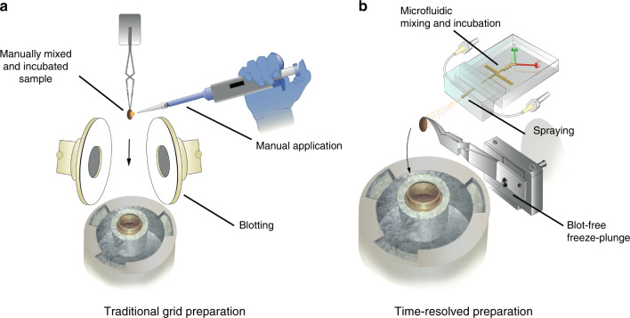
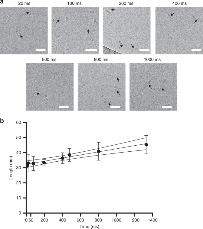
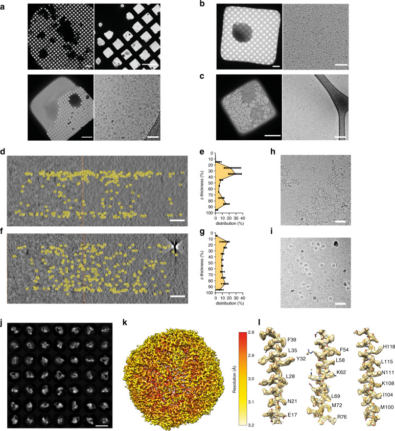
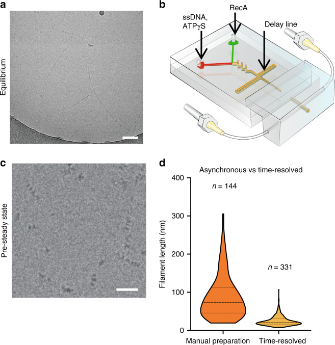
Mechanistic understanding of biochemical reactions requires structural and kinetic characterization of the underlying chemical processes. However, no single experimental technique can provide this information in a broadly applicable manner and thus structural studies of static macromolecules are often complemented by biophysical analysis. Moreover, the common strategy of utilizing mutants or crosslinking probes to stabilize intermediates is prone to trapping off-pathway artefacts and precludes determining the order of molecular events. Here we report a time-resolved sample preparation method for cryo-electron microscopy (trEM) using a modular microfluidic device, featuring a 3D-mixing unit and variable delay lines that enables automated, fast, and blot-free sample vitrification. This approach not only preserves high-resolution structural detail but also substantially improves sample integrity and protein distribution across the vitreous ice. We validate the method by visualising reaction intermediates of early RecA filament growth across three orders of magnitude on sub-second timescales. The trEM method reported here is versatile, reproducible, and readily adaptable to a broad spectrum of fundamental questions in biology.
生化反应的机制理解需要对潜在化学过程进行结构和动力学表征。然而,没有单一的实验技术可以以广泛适用的方式提供这些信息,因此静态大分子的结构研究通常需要生物物理分析的补充。此外,利用突变体或交联探针来稳定中间产物的常见策略容易捕获偏离途径的伪影,并且无法确定分子事件的顺序。在这里,我们报告了一种使用模块化微流控装置的用于冷冻电子显微镜(trEM)的时间分辨样品制备方法,该装置具有 3D 混合单元和可变延迟线,可实现自动化、快速且无斑点的样品玻璃化。这种方法不仅保留了高分辨率的结构细节,而且大大提高了样品的完整性和蛋白质在玻璃态冰中的分布。我们通过在亚秒时间尺度上可视化早期 RecA 丝生长的反应中间体跨越三个数量级来验证该方法。这里报道的 trEM 方法具有多功能性、可重复性,并且易于适应生物学中广泛的基本问题。