GKT School of Medical Education, King's College London, London, UK.
School of Biomedical Engineering and Imaging Sciences, King's College London, London, UK.
Int J Comput Assist Radiol Surg. 2020 Sep;15(9):1445-1455. doi: 10.1007/s11548-020-02222-y. Epub 2020 Jul 16.
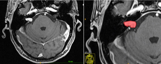
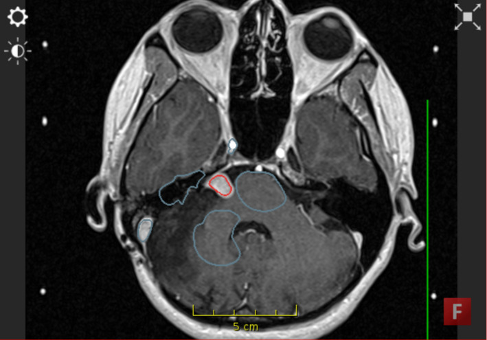
Management of vestibular schwannoma (VS) is based on tumour size as observed on T1 MRI scans with contrast agent injection. The current clinical practice is to measure the diameter of the tumour in its largest dimension. It has been shown that volumetric measurement is more accurate and more reliable as a measure of VS size. The reference approach to achieve such volumetry is to manually segment the tumour, which is a time intensive task. We suggest that semi-automated segmentation may be a clinically applicable solution to this problem and that it could replace linear measurements as the clinical standard.
Using high-quality software available for academic purposes, we ran a comparative study of manual versus semi-automated segmentation of VS on MRI with 5 clinicians and scientists. We gathered both quantitative and qualitative data to compare the two approaches; including segmentation time, segmentation effort and segmentation accuracy.
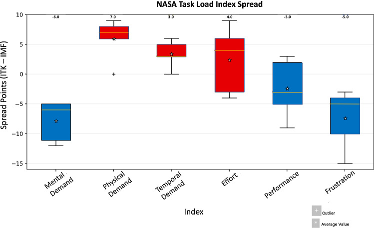
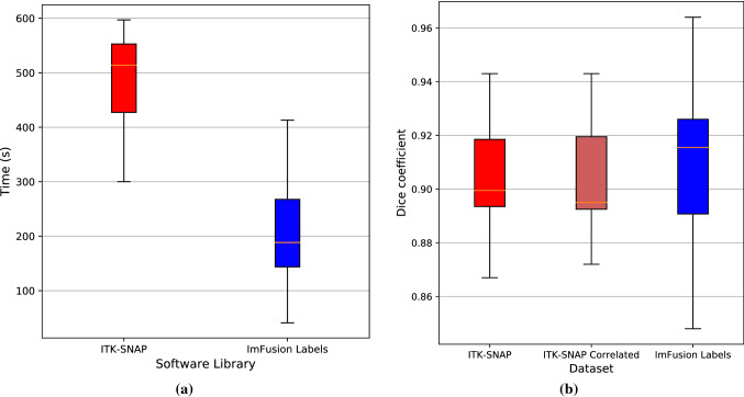
We found that the selected semi-automated segmentation approach is significantly faster (167 s vs 479 s, [Formula: see text]), less temporally and physically demanding and has approximately equal performance when compared with manual segmentation, with some improvements in accuracy. There were some limitations, including algorithmic unpredictability and error, which produced more frustration and increased mental effort in comparison with manual segmentation.
We suggest that semi-automated segmentation could be applied clinically for volumetric measurement of VS on MRI. In future, the generic software could be refined for use specifically for VS segmentation, thereby improving accuracy.
听神经鞘瘤(VS)的管理基于 T1 MRI 扫描时注射造影剂后观察到的肿瘤大小。目前的临床实践是测量肿瘤在最大维度上的直径。已经证明,体积测量作为 VS 大小的衡量标准更准确、更可靠。实现这种体积测量的参考方法是手动分割肿瘤,但这是一项耗时的任务。我们建议,半自动分割可能是解决此问题的一种临床适用的解决方案,并且它可以替代线性测量作为临床标准。
我们使用了高质量的、可供学术使用的软件,让 5 名临床医生和科学家对 MRI 上的 VS 手动分割与半自动分割进行了对比研究。我们收集了定量和定性数据来比较这两种方法;包括分割时间、分割难度和分割准确性。
我们发现,所选的半自动分割方法明显更快(167s 与 479s,[公式:见正文]),耗时和体力要求更低,与手动分割相比具有相当的性能,准确性略有提高。存在一些限制,包括算法的不可预测性和错误,与手动分割相比,这些错误会产生更多的挫折感和更大的精神压力。
我们建议,可以将半自动分割应用于 MRI 上 VS 的体积测量的临床实践中。在未来,通用软件可以进行细化,专门用于 VS 分割,从而提高准确性。