Sun Xiaolei, Li Xueping, Qi Hongzhao, Hou Xin, Zhao Jin, Yuan Xubo, Ma Xinlong
Tianjin Key Laboratory of Composite and Functional Materials, School of Materials Science and Engineering, Tianjin University, Tianjin, 300072, China.
Department of Orthopaedics, Tianjin Hospital, Tianjin, 300211, China.
J Orthop Translat. 2020 May 19;24:76-87. doi: 10.1016/j.jot.2020.04.007. eCollection 2020 Sep.
The healing of osteoporotic fractures in the elderly patients is a difficult clinical problem. Currently, based on the internal fixation of fractures, the available drug treatments mainly focus on either inhibiting osteoclast function, such as bisphosphonate, calcitonin, oestrogen or promoting osteogenesis, such as parathyroid hormones. However, the availability of current antiosteoporotic drugs in promoting osteoporotic fracture healing is limited. The objective of the present study was to investigate the ability of the MiR-21/nanocapsule to enhance the early bone repair of osteoporotic fractures.
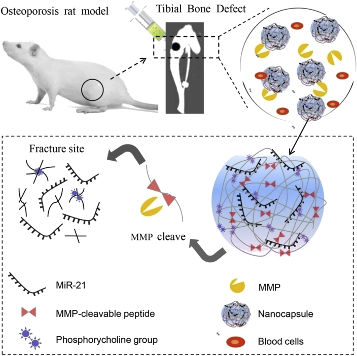
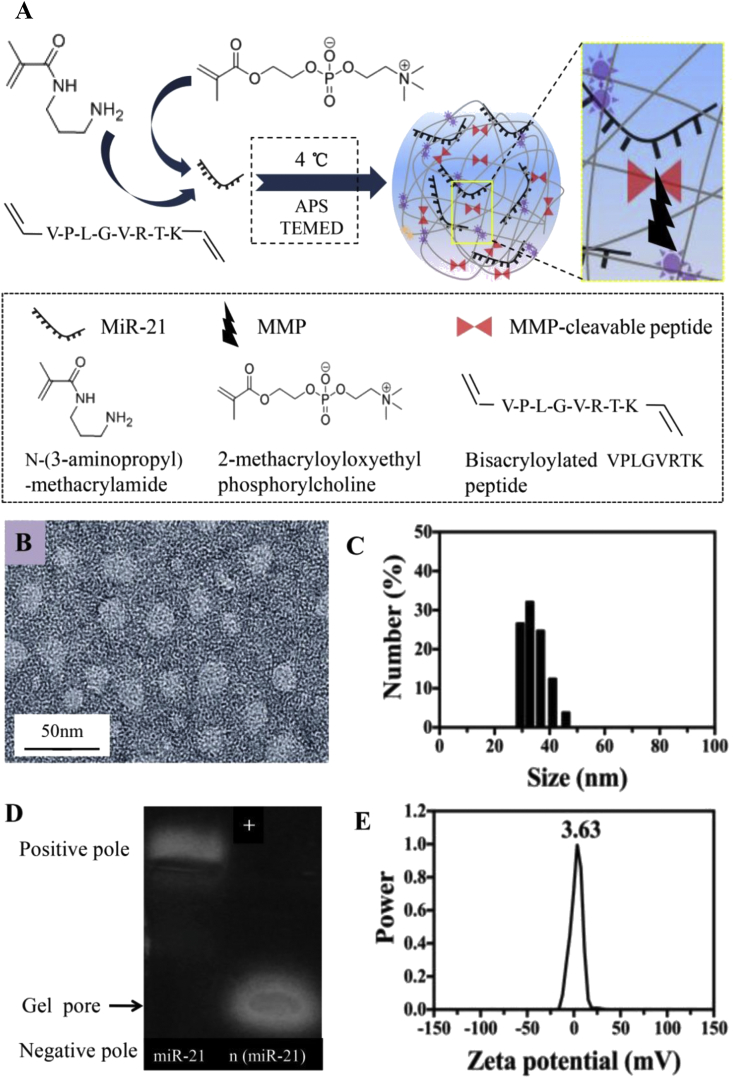
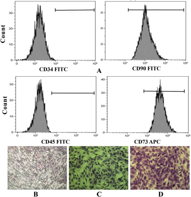
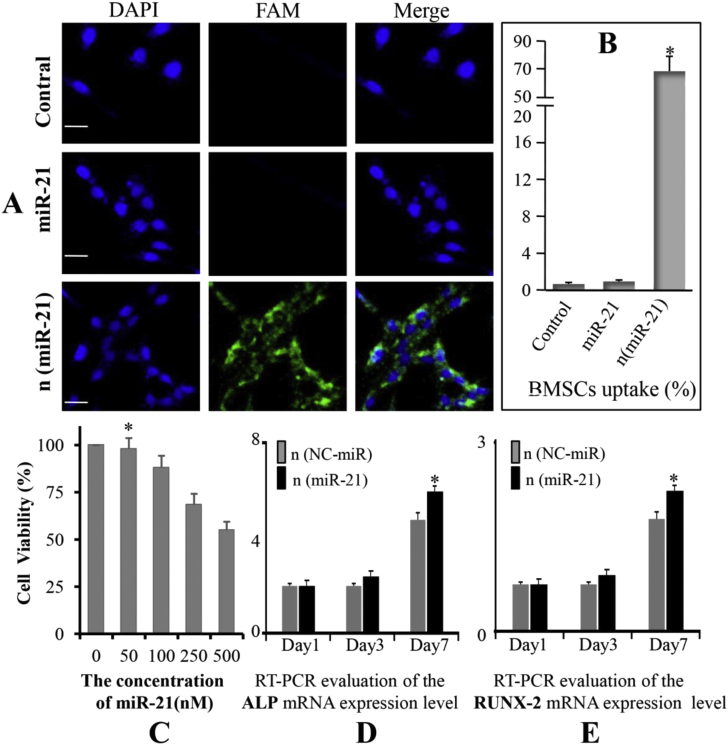
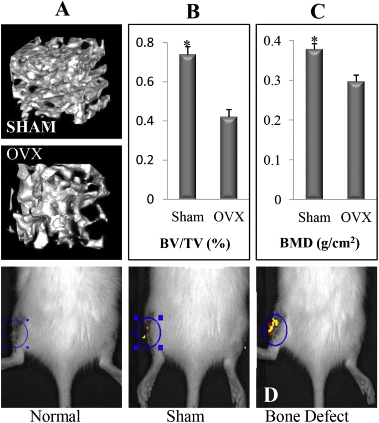
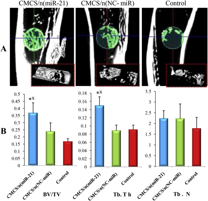
Based on the presence of matrix metalloproteinases that are overexpressed at the fracture site, we designed the matrix metalloproteinase-sensitive nanocapsules which were formed by in situ free radical polymerisation on the surface of MiR-21 with 2-(methacryloyloxy) ethyl phosphorylcholine and the bisacryloylated VPLGVRTK peptide. The MiR-21/nanocapsule [n (miR-21)] and O-carboxymethyl chitosan (CMCS) were mixed until they formed a gel-like material [CMCS/n (miR-21)] with good fluidity and injectability. Thirty elderly Sprague Dawley (SD) rats (female, 14-month-old, 380 ± 10 g) were subjected to bilateral removal of the ovaries (ovariectomised). All rats were subjected to bilateral bone defects (2 mm diameter) of the proximal tibia and randomly divided into three groups (groups A, B, and C): separately injected with CMCS/n (miR-21), CMCS/n (NC-miR), and saline. Micro-computed tomography (CT) imaging was performed to evaluate newly formed bone volume and connectivity. Nondecalcified histology and toluidine blue staining were performed to measure the effects of CMCS/n (miR-21) on bone repair. In vitro, the effect of n (miR-21) on osteogenic differentiation to bone marrow mesenchymal stem cells (BMSCs) which derived from the ovariectomised rat model was observed.
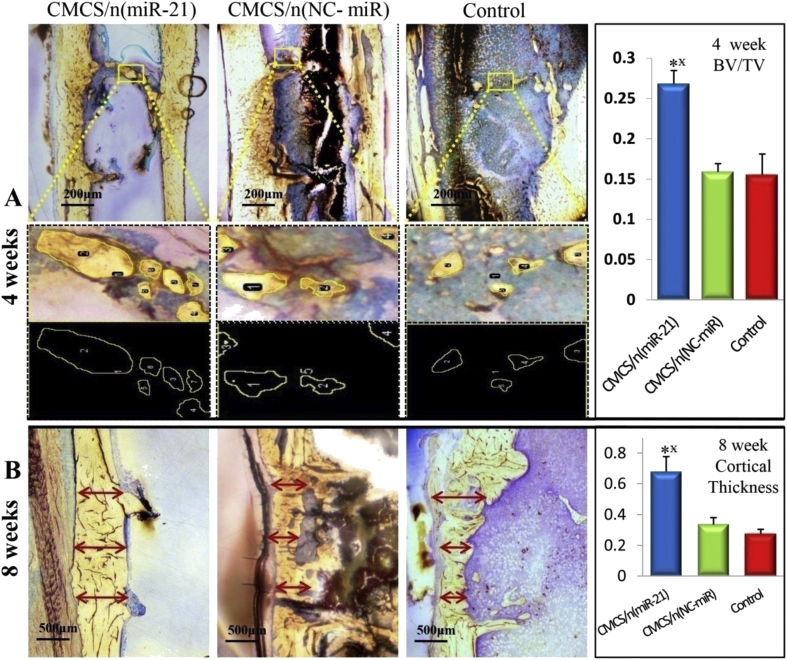
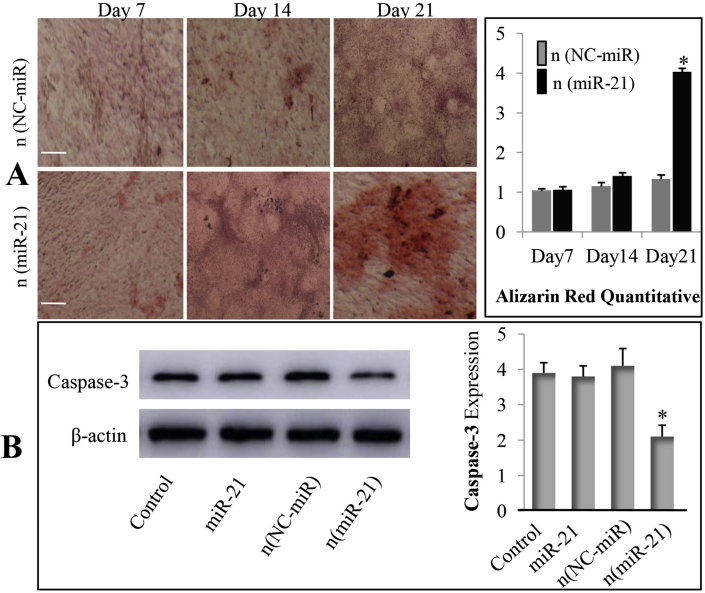
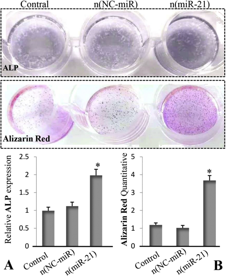
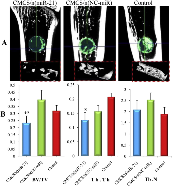
The morphology of n (miR-21) was a regular spherical nanocapsule with a uniform small size (25-35 nm). The results confirmed that n (miR-21) could be efficiently phagocytosed by BMSCs and released in the cytoplasm to promote osteogenesis. The expression level of alkaline phosphatase and Runt-related transcription factor 2 mRNA in the n (miR-21) group was higher than that in the n (NC-miR) group. Animal experiments proved that CMCS/n (miR-21) produced better bone repair compared with the CMCS/n (NC-miR) group in the early stages of fracture healing at 4 weeks. In the late stage of fracture healing (8 weeks), micro-CT quantitative analysis showed that the new bone trabeculae in the CMCS/n (miR-21) group has decreased compared with the CMCS/n (NC-miR) group. In the CMCS/n (miR-21) group, the new cancellous bone had been absorbed, and the process of bone healing was almost completed. In contrast, the new bone in the CMCS/n (NC-miR) and the control groups was still in the healing process.
The cytological tests confirmed that n (miR-21) can promote osteogenic differentiation of BMSCs derived from the osteoporosis rat model. Furthermore, the results of animal tests demonstrated that local injection of CMCS/n (miR-21) promoted the early healing of osteoporotic bone defects. Consequently CMCS/n (miR-21) promoted the bone repair process to enter the moulding phase earlier.
CMCS/n (miR-21) can be widely applied to elderly patients with osteoporotic fractures. This method can help patients with osteoporotic fractures recover earlier and avoid serious complications. It provides a potential approach for the clinical treatment of osteoporotic fractures in the elderly.
老年患者骨质疏松性骨折的愈合是一个棘手的临床问题。目前,在骨折内固定的基础上,现有的药物治疗主要集中在抑制破骨细胞功能,如双膦酸盐、降钙素、雌激素,或促进成骨,如甲状旁腺激素。然而,目前的抗骨质疏松药物在促进骨质疏松性骨折愈合方面的作用有限。本研究的目的是探讨MiR-21/纳米胶囊促进骨质疏松性骨折早期骨修复的能力。
基于骨折部位过表达的基质金属蛋白酶,我们设计了基质金属蛋白酶敏感的纳米胶囊,其通过在MiR-21表面用2-(甲基丙烯酰氧基)乙基磷酰胆碱和双丙烯酰化的VPLGVRTK肽进行原位自由基聚合形成。将MiR-21/纳米胶囊[n(miR-21)]与O-羧甲基壳聚糖(CMCS)混合,直至形成具有良好流动性和可注射性的凝胶状材料[CMCS/n(miR-21)]。30只老年Sprague Dawley(SD)大鼠(雌性,14月龄,380±10g)接受双侧卵巢切除(去卵巢)。所有大鼠均接受双侧胫骨近端骨缺损(直径2mm),并随机分为三组(A组、B组和C组):分别注射CMCS/n(miR-21)、CMCS/n(NC-miR)和生理盐水。进行微型计算机断层扫描(CT)成像以评估新形成的骨体积和连通性。进行非脱钙组织学和甲苯胺蓝染色以测量CMCS/n(miR-21)对骨修复的影响。在体外,观察n(miR-21)对去卵巢大鼠模型来源的骨髓间充质干细胞(BMSCs)成骨分化的影响。
n(miR-21)的形态为规则的球形纳米胶囊,尺寸均匀较小(25-35nm)。结果证实n(miR-21)可被BMSCs有效吞噬并在细胞质中释放以促进成骨。n(miR-21)组碱性磷酸酶和Runt相关转录因子2 mRNA的表达水平高于n(NC-miR)组。动物实验证明,在骨折愈合早期4周时,与CMCS/n(NC-miR)组相比,CMCS/n(miR-21)产生了更好的骨修复效果。在骨折愈合后期(8周),微型CT定量分析显示,CMCS/n(miR-21)组的新骨小梁比CMCS/n(NC-miR)组减少。在CMCS/n(miR-21)组中,新的松质骨已被吸收,骨愈合过程几乎完成。相比之下,CMCS/n(NC-miR)组和对照组的新骨仍处于愈合过程中。
细胞学试验证实n(miR-21)可促进骨质疏松大鼠模型来源的BMSCs的成骨分化。此外,动物试验结果表明,局部注射CMCS/n(miR-21)可促进骨质疏松性骨缺损的早期愈合。因此,CMCS/n(miR-21)促进骨修复过程更早进入塑形期。
CMCS/n(miR-21)可广泛应用于老年骨质疏松性骨折患者。该方法可帮助骨质疏松性骨折患者更早康复,避免严重并发症。它为老年骨质疏松性骨折的临床治疗提供了一种潜在的方法。