Harrison Judith R, Bhatia Sanchita, Tan Zhao Xuan, Mirza-Davies Anastasia, Benkert Hannah, Tax Chantal M W, Jones Derek K
Cardiff University Brain Research Imaging Centre (CUBRIC), Maindy Road, Cardiff CF24 4HQ, UK.
Cardiff University School of Medicine, University Hospital of Wales, Heath Park, Cardiff CF14 4XN, UK.
Neuroimage Clin. 2020;27:102359. doi: 10.1016/j.nicl.2020.102359. Epub 2020 Jul 22.
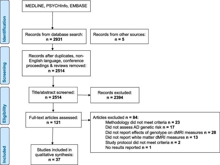
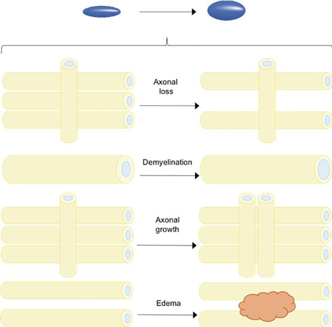
Diffusion magnetic resonance imaging (dMRI) is an imaging technique which probes the random motion of water molecules in tissues and has been widely applied to investigate changes in white matter microstructure in Alzheimer's Disease. This paper aims to systematically review studies that examined the effect of Alzheimer's risk genes on white matter microstructure. We assimilated findings from 37 studies and reviewed their diffusion pre-processing and analysis methods. Most studies estimate the diffusion tensor (DT) and compare derived quantitative measures such as fractional anisotropy and mean diffusivity between groups. Those with increased AD genetic risk are associated with reduced anisotropy and increased diffusivity across the brain, most notably the temporal and frontal lobes, cingulum and corpus callosum. Structural abnormalities are most evident amongst those with established Alzheimer's Disease. Recent studies employ signal representations and analysis frameworks beyond DT MRI but show that dMRI overall lacks specificity to disease pathology. However, as the field advances, these techniques may prove useful in pre-symptomatic diagnosis or staging of Alzheimer's disease.
扩散磁共振成像(dMRI)是一种探测组织中水分子随机运动的成像技术,已被广泛应用于研究阿尔茨海默病白质微观结构的变化。本文旨在系统综述研究阿尔茨海默病风险基因对白质微观结构影响的相关研究。我们汇总了37项研究的结果,并回顾了它们的扩散预处理和分析方法。大多数研究估计扩散张量(DT),并比较不同组之间导出的定量指标,如分数各向异性和平均扩散率。阿尔茨海默病遗传风险增加的人群与全脑各向异性降低和扩散率增加有关,最明显的是颞叶、额叶、扣带回和胼胝体。结构异常在已确诊的阿尔茨海默病患者中最为明显。最近的研究采用了超越DT MRI的信号表示和分析框架,但表明dMRI总体上对疾病病理缺乏特异性。然而,随着该领域的发展,这些技术可能在阿尔茨海默病的症状前诊断或分期中证明有用。