Feng Di, Zhou Huanping, Jin Xiaohong, Wei Juan, Zhang Qingqing, Gu Yang, Zhang Pengcheng, Yang Hao, Song Jiangang, Shi Xuan, Lv Xin
Department of Anesthesiology, Shanghai Pulmonary Hospital, Tongji University School of Medicine, Shanghai 200433, China.
Department of Anesthesiology, The Second Affiliated Hospital of Nanchang University, Nanchang 330006, China.
Evid Based Complement Alternat Med. 2020 Jul 22;2020:4594631. doi: 10.1155/2020/4594631. eCollection 2020.
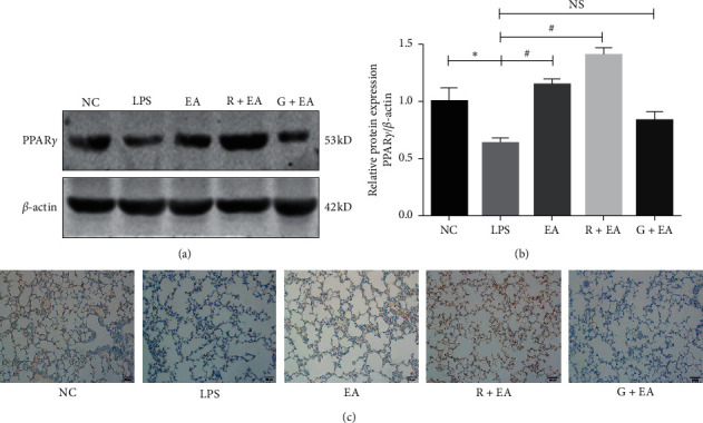
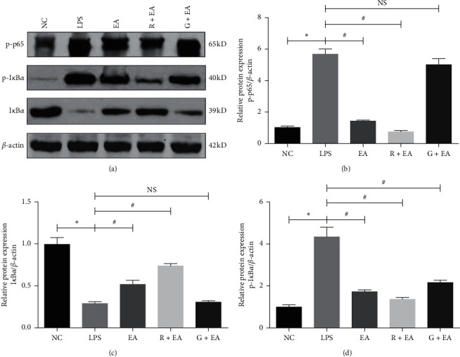
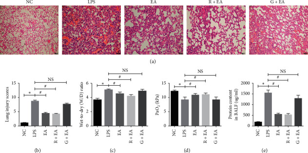
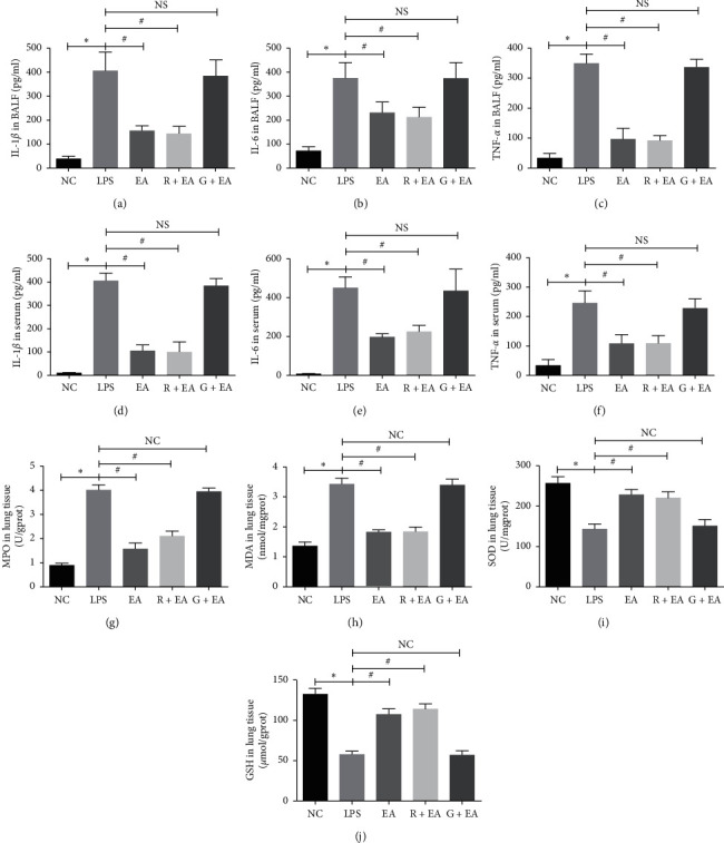
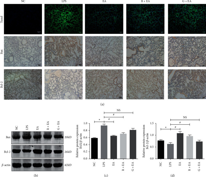
Electroacupuncture (EA) is reported to possess anti-inflammatory properties and has beneficial effects on acute respiratory distress syndrome (ARDS). However, the underlying mechanisms of the effects of EA on ARDS remain unclear. This study aims to investigate the protective effect of EA on LPS-induced ARDS. In this study, Sprague-Dawley male rats were treated with EA at Hegu (LI4) for 45 minutes before LPS instillation (0.4 mg/kg, 100 ul). H&E staining, wet-to-dry weight (W/D) ratio, PaO, and protein content in BALF were employed to determine the function of lung tissues. Inflammatory cytokines in serum and BALF were detected by enzyme-linked immunoassay assay (ELISA). The levels of oxidative stress markers were detected to determine the oxidative stress status. Cell apoptosis was observed by terminal deoxynucleotidyl transferase-mediated dUTP nick-end labeling (TUNEL) staining and western blot. Here, we found that EA pretreatment effectively alleviated lung pathological damage. Moreover, EA suppressed the oxidative stress damage by upregulating glutathione and superoxide dismutase and downregulating malondialdehyde. EA pretreatment also regulated apoptosis-related proteins, such as Bax and Bcl-2. We found that peroxisome proliferators-activated receptors (PPAR) play a critical role during ARDS, EA up-regulated the expression of PPAR, which inhibited the activation of nuclear factor-kappa B (NF-B) and decreased the inflammatory cytokines (interleukin-1, interleukin-6, and tumor necrosis factor-). When rats were treated with GW9662, a selective PPAR antagonist, these effects of EA were reversed. Our study demonstrated that EA pretreatment had a beneficial effect on LPS-induced ARDS in rats by anti-inflammatory, antioxidative, and antiapoptotic properties which was regulated via PPAR/NF-B signaling pathway.
据报道,电针(EA)具有抗炎特性,对急性呼吸窘迫综合征(ARDS)有有益作用。然而,EA对ARDS作用的潜在机制仍不清楚。本研究旨在探讨EA对脂多糖(LPS)诱导的ARDS的保护作用。在本研究中,将Sprague-Dawley雄性大鼠在滴注LPS(0.4mg/kg,100μl)前于合谷穴(LI4)进行45分钟的电针治疗。采用苏木精-伊红(H&E)染色、湿重与干重(W/D)比值、动脉血氧分压(PaO)以及支气管肺泡灌洗液(BALF)中的蛋白质含量来测定肺组织功能。通过酶联免疫吸附测定(ELISA)检测血清和BALF中的炎性细胞因子。检测氧化应激标志物水平以确定氧化应激状态。通过末端脱氧核苷酸转移酶介导的dUTP缺口末端标记(TUNEL)染色和蛋白质印迹法观察细胞凋亡。在此,我们发现电针预处理有效减轻了肺病理损伤。此外,电针通过上调谷胱甘肽和超氧化物歧化酶并下调丙二醛来抑制氧化应激损伤。电针预处理还调节了凋亡相关蛋白,如Bax和Bcl-2。我们发现过氧化物酶体增殖物激活受体(PPAR)在ARDS过程中起关键作用,电针上调了PPAR的表达,这抑制了核因子-κB(NF-κB)的激活并减少了炎性细胞因子(白细胞介素-1、白细胞介素-6和肿瘤坏死因子-α)。当用选择性PPAR拮抗剂GW9662处理大鼠时,电针的这些作用被逆转。我们的研究表明,电针预处理通过抗炎、抗氧化和抗凋亡特性对LPS诱导的大鼠ARDS具有有益作用,其通过PPAR/NF-κB信号通路进行调节。