• 文献检索
  • 文档翻译
  • 深度研究
  • 学术资讯
  • Suppr Zotero 插件Zotero 插件
  • 邀请有礼
  • 套餐&价格
  • 历史记录
应用&插件
Suppr Zotero 插件Zotero 插件浏览器插件Mac 客户端Windows 客户端微信小程序
定价
高级版会员购买积分包购买API积分包
服务
文献检索文档翻译深度研究API 文档MCP 服务
关于我们
关于 Suppr公司介绍联系我们用户协议隐私条款
关注我们

Suppr 超能文献

核心技术专利:CN118964589B侵权必究
粤ICP备2023148730 号-1Suppr @ 2026

文献检索

告别复杂PubMed语法,用中文像聊天一样搜索,搜遍4000万医学文献。AI智能推荐,让科研检索更轻松。

立即免费搜索

文件翻译

保留排版,准确专业,支持PDF/Word/PPT等文件格式,支持 12+语言互译。

免费翻译文档

深度研究

AI帮你快速写综述,25分钟生成高质量综述,智能提取关键信息,辅助科研写作。

立即免费体验

光学微血管成像揭示了在胡须刺激期间小鼠皮层桶状皮层中的时变和深度分辨的血液动力学变化。

Optical microangiography reveals temporal and depth-resolved hemodynamic change in mouse barrel cortex during whisker stimulation.

机构信息

University of Washington, Department of Bioengineering, Seattle, Washington, United States.

出版信息

J Biomed Opt. 2020 Sep;25(9). doi: 10.1117/1.JBO.25.9.096005.

DOI:10.1117/1.JBO.25.9.096005
PMID:32945154
原文链接:https://pmc.ncbi.nlm.nih.gov/articles/PMC7495356/
Abstract

SIGNIFICANCE

Cerebral blood flow (CBF) regulation at neurovascular coupling (NVC) plays an important role in normal brain functioning to support oxygen delivery to activating neurons. Therefore, studying the mechanisms of CBF adjustment is crucial for the improved understanding of brain activity.

AIM

We investigated the temporal profile of hemodynamic signal change in mouse cortex caused by neural activation and its variation over cortical depth.

APPROACH

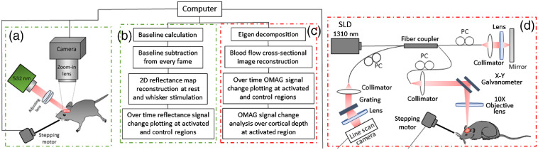
Following the cranial window surgery, intrinsic optical signal imaging (IOSI) was used to spatially locate the activated region in mouse cortex during whisker stimulation. Optical microangiography (OMAG), the functional extension of optical coherence tomography, was applied to image the activated and control regions identified by IOSI. Temporal profiles of hemodynamic response signals obtained by IOSI and OMAG were compared, and OMAG signal was analyzed over cortical layers.

RESULTS

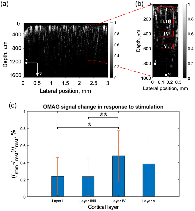
Our results showed that the hemodynamic response to neural activity revealed by blood flow change signal signal through IOSI is slower than that observed by OMAG signal. OMAG also indicated the laminar variation of the response over cortical depth, showing the largest response in cortical layer IV.

CONCLUSIONS

Overall, we demonstrated the development and application of dual-modality imaging system composed of IOSI and OMAG, which may have potential to enable the future investigations of depth-resolved CBF and to provide the insights of hemodynamic events associated with the NVC.

摘要

意义

神经血管耦合(NVC)时的脑血流(CBF)调节在正常大脑功能中起着重要作用,以支持向激活神经元供氧。因此,研究 CBF 调节的机制对于更好地理解大脑活动至关重要。

目的

我们研究了由神经激活引起的小鼠皮层血流动力学信号变化的时间分布及其在皮层深度上的变化。

方法

在颅窗手术后,使用固有光学信号成像(IOSI)在胡须刺激期间在小鼠皮层中空间定位激活区域。光学微血管造影(OMAG)是光相干断层扫描的功能扩展,用于对 IOSI 识别的激活和对照区域进行成像。比较了 IOSI 和 OMAG 获得的血流动力学响应信号的时间分布,并对皮层层进行了 OMAG 信号分析。

结果

我们的结果表明,通过 IOSI 测量的血流变化信号揭示的神经活动引起的血液动力学反应比 OMAG 信号观察到的要慢。OMAG 还表明,响应在皮层深度上的分层变化,在皮层 IV 层中显示出最大的响应。

结论

总体而言,我们展示了由 IOSI 和 OMAG 组成的双模成像系统的开发和应用,这可能为未来对深度分辨 CBF 的研究提供潜力,并提供与 NVC 相关的血液动力学事件的见解。

https://cdn.ncbi.nlm.nih.gov/pmc/blobs/5fc9/7495356/8318f41d8f7d/JBO-025-096005-g004.jpg
https://cdn.ncbi.nlm.nih.gov/pmc/blobs/5fc9/7495356/6c555847b8ab/JBO-025-096005-g001.jpg
https://cdn.ncbi.nlm.nih.gov/pmc/blobs/5fc9/7495356/0bf6e5661f4a/JBO-025-096005-g002.jpg
https://cdn.ncbi.nlm.nih.gov/pmc/blobs/5fc9/7495356/f828cba7d3b7/JBO-025-096005-g003.jpg
https://cdn.ncbi.nlm.nih.gov/pmc/blobs/5fc9/7495356/8318f41d8f7d/JBO-025-096005-g004.jpg

相似文献

1
Optical microangiography reveals temporal and depth-resolved hemodynamic change in mouse barrel cortex during whisker stimulation.光学微血管成像揭示了在胡须刺激期间小鼠皮层桶状皮层中的时变和深度分辨的血液动力学变化。
J Biomed Opt. 2020 Sep;25(9). doi: 10.1117/1.JBO.25.9.096005.
2
Functional evaluation of hemodynamic response during neural activation using optical microangiography integrated with dual-wavelength laser speckle imaging.采用双波长激光散斑成像技术集成的光学微脉管造影术对神经激活过程中的血液动力学反应进行功能评估。
J Biomed Opt. 2014 Feb;19(2):026013. doi: 10.1117/1.JBO.19.2.026013.
3
Impact of Altered Cholinergic Tones on the Neurovascular Coupling Response to Whisker Stimulation.胆碱能张力改变对触须刺激神经血管耦合反应的影响。
J Neurosci. 2017 Feb 8;37(6):1518-1531. doi: 10.1523/JNEUROSCI.1784-16.2016. Epub 2017 Jan 9.
4
Differences in cerebral blood vasculature and flow in awake and anesthetized mouse cortex revealed by quantitative optical coherence tomography angiography.定量光学相干断层扫描血管造影术揭示清醒和麻醉小鼠皮层中脑血流血管的差异。
J Neurosci Methods. 2021 Apr 1;353:109094. doi: 10.1016/j.jneumeth.2021.109094. Epub 2021 Feb 5.
5
Procedure and protocols for optical imaging of cerebral blood flow and hemodynamics in awake mice.清醒小鼠脑血流和血流动力学光学成像的程序与方案
Biomed Opt Express. 2020 May 26;11(6):3288-3300. doi: 10.1364/BOE.394649. eCollection 2020 Jun 1.
6
Optical Coherence Tomography angiography reveals laminar microvascular hemodynamics in the rat somatosensory cortex during activation.光学相干断层扫描血管造影术揭示了激活过程中大鼠体感皮层的层流微血管血流动力学。
Neuroimage. 2014 Nov 15;102 Pt 2(0 2):393-406. doi: 10.1016/j.neuroimage.2014.08.004. Epub 2014 Aug 8.
7
Development of new optical imaging systems of oxygen metabolism and simultaneous measurement in hemodynamic changes using awake mice.利用清醒小鼠开发新的氧代谢光学成像系统并同步测量血流动力学变化。
J Neurosci Methods. 2014 Nov 30;237:9-15. doi: 10.1016/j.jneumeth.2014.08.022. Epub 2014 Sep 1.
8
Measurement and visualization of stimulus-evoked tissue dynamics in mouse barrel cortex using phase-sensitive optical coherence tomography.使用相敏光学相干断层扫描技术测量和可视化小鼠桶状皮层中刺激诱发的组织动力学。
Biomed Opt Express. 2020 Jan 9;11(2):699-710. doi: 10.1364/BOE.381332. eCollection 2020 Feb 1.
9
Quantitative In Vivo Imaging of Tissue Absorption, Scattering, and Hemoglobin Concentration in Rat Cortex Using Spatially Modulated Structured Light使用空间调制结构光对大鼠皮层组织吸收、散射和血红蛋白浓度进行定量体内成像
10
In Vivo Observations of Rapid Scattered Light Changes Associated with Neurophysiological Activity与神经生理活动相关的快速散射光变化的体内观察

引用本文的文献

1
Optical coherence tomography (OCT) and OCT angiography: Technological development and applications in brain science.光学相干断层扫描(OCT)与OCT血管造影:技术发展及其在脑科学中的应用
Theranostics. 2025 Jan 1;15(1):122-140. doi: 10.7150/thno.97192. eCollection 2025.
2
Neurophotonic tools for microscopic measurements and manipulation: status report.用于微观测量和操作的神经光子学工具:现状报告。
Neurophotonics. 2022 Jan;9(Suppl 1):013001. doi: 10.1117/1.NPh.9.S1.013001. Epub 2022 Apr 27.

本文引用的文献

1
Procedure and protocols for optical imaging of cerebral blood flow and hemodynamics in awake mice.清醒小鼠脑血流和血流动力学光学成像的程序与方案
Biomed Opt Express. 2020 May 26;11(6):3288-3300. doi: 10.1364/BOE.394649. eCollection 2020 Jun 1.
2
A noninvasive imaging and measurement using optical coherence tomography angiography for the assessment of gingiva: An in vivo study.一项使用光学相干断层扫描血管造影术对牙龈进行评估的无创成像与测量:一项体内研究。
J Biophotonics. 2018 Dec;11(12):e201800242. doi: 10.1002/jbio.201800242. Epub 2018 Sep 5.
3
Capillary flow homogenization during functional activation revealed by optical coherence tomography angiography based capillary velocimetry.
基于光相干断层扫描血管造影的毛细血管速度测量术显示功能激活过程中的毛细血管流动均化。
Sci Rep. 2018 Mar 7;8(1):4107. doi: 10.1038/s41598-018-22513-4.
4
Optical coherence tomography angiography of normal skin and inflammatory dermatologic conditions.正常皮肤和炎症性皮肤病的光学相干断层扫描血管造影
Lasers Surg Med. 2018 Mar;50(3):183-193. doi: 10.1002/lsm.22788. Epub 2018 Jan 22.
5
Intrinsic signal optical imaging of visual brain activity: Tracking of fast cortical dynamics.视觉脑活动的内在信号光学成像:快速皮层动力学的追踪
Neuroimage. 2017 Mar 1;148:160-168. doi: 10.1016/j.neuroimage.2017.01.006. Epub 2017 Jan 4.
6
Characterizing relationship between optical microangiography signals and capillary flow using microfluidic channels.利用微流控通道表征光学微血管造影信号与毛细血管流动之间的关系。
Biomed Opt Express. 2016 Jun 20;7(7):2709-28. doi: 10.1364/BOE.7.002709. eCollection 2016 Jul 1.
7
Effect of electrical forepaw stimulation on capillary transit-time heterogeneity (CTH).前爪电刺激对毛细血管传输时间异质性(CTH)的影响。
J Cereb Blood Flow Metab. 2016 Dec;36(12):2072-2086. doi: 10.1177/0271678X16631560. Epub 2016 Feb 8.
8
Laminar microvascular transit time distribution in the mouse somatosensory cortex revealed by Dynamic Contrast Optical Coherence Tomography.动态对比光学相干断层扫描揭示小鼠体感皮层中的层流微血管传输时间分布
Neuroimage. 2016 Jan 15;125:350-362. doi: 10.1016/j.neuroimage.2015.10.017. Epub 2015 Oct 20.
9
Vasodynamics of pial and penetrating arterioles in relation to arteriolo-arteriolar anastomosis after focal stroke.皮质和穿透性小动脉的血管动力学与局灶性脑卒中后小动脉-小动脉吻合的关系。
Neurophotonics. 2015 Apr;2(2):025006. doi: 10.1117/1.NPh.2.2.025006. Epub 2015 Jun 12.
10
Application of thinned-skull cranial window to mouse cerebral blood flow imaging using optical microangiography.薄颅骨脑窗在小鼠脑血流光学微血管造影成像中的应用。
PLoS One. 2014 Nov 26;9(11):e113658. doi: 10.1371/journal.pone.0113658. eCollection 2014.