Srinivasan Vivek J, Radhakrishnan Harsha
Department of Biomedical Engineering, University of California at Davis, 451 E. Health Sciences Dr. GBSF 2303, Davis, CA 95616, USA.
Department of Biomedical Engineering, University of California at Davis, 451 E. Health Sciences Dr. GBSF 2303, Davis, CA 95616, USA.
Neuroimage. 2014 Nov 15;102 Pt 2(0 2):393-406. doi: 10.1016/j.neuroimage.2014.08.004. Epub 2014 Aug 8.
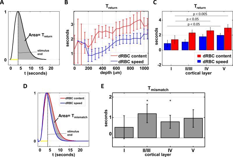
The BOLD (blood-oxygen-level dependent) fMRI (functional Magnetic Resonance Imaging) signal is shaped, in part, by changes in red blood cell (RBC) content and flow across vascular compartments over time. These complex dynamics have been challenging to characterize directly due to a lack of appropriate imaging modalities. In this study, making use of infrared light scattering from RBCs, depth-resolved Optical Coherence Tomography (OCT) angiography was applied to image laminar functional hyperemia in the rat somatosensory cortex. After defining and validating depth-specific metrics for changes in RBC content and speed, laminar hemodynamic responses in microvasculature up to cortical depths of >1mm were measured during a forepaw stimulus. The results provide a comprehensive picture of when and where changes in RBC content and speed occur during and immediately following cortical activation. In summary, the earliest and largest microvascular RBC content changes occurred in the middle cortical layers, while post-stimulus undershoots were most prominent superficially. These laminar variations in positive and negative responses paralleled known distributions of excitatory and inhibitory synapses, suggesting neuronal underpinnings. Additionally, the RBC speed response consistently returned to baseline more promptly than RBC content after the stimulus across cortical layers, supporting a "flow-volume mismatch" of hemodynamic origin.
血氧水平依赖性功能磁共振成像(BOLD-fMRI)信号部分由红细胞(RBC)含量的变化以及随着时间推移血管腔室中血流的变化所塑造。由于缺乏合适的成像方式,这些复杂的动力学过程一直难以直接表征。在本研究中,利用红细胞的红外光散射,将深度分辨光学相干断层扫描(OCT)血管造影应用于大鼠体感皮层的层流功能性充血成像。在定义并验证了红细胞含量和速度变化的深度特异性指标后,在前爪刺激期间测量了直至皮层深度>1mm的微脉管系统中的层流血流动力学反应。结果提供了一幅在皮层激活期间及之后红细胞含量和速度变化发生的时间和位置的全面图景。总之,最早且最大的微血管红细胞含量变化发生在皮层中间层,而刺激后的负向波在最表层最为显著。这些正负反应中的层流变化与已知的兴奋性和抑制性突触分布平行,提示存在神经元基础。此外,刺激后跨皮层各层红细胞速度反应始终比红细胞含量更快地恢复到基线水平,支持了血流动力学起源的“流量-容积不匹配”。