Norwegian Center for Mental Disorders Research (NORMENT), Division of Mental Health and Addiction, Oslo University Hospital, Oslo, Norway.
Norwegian Center for Mental Disorders Research (NORMENT), Institute of Clinical Medicine, University of Oslo, Oslo, Norway.
Hum Brain Mapp. 2022 Jan;43(1):373-384. doi: 10.1002/hbm.25212. Epub 2020 Oct 5.
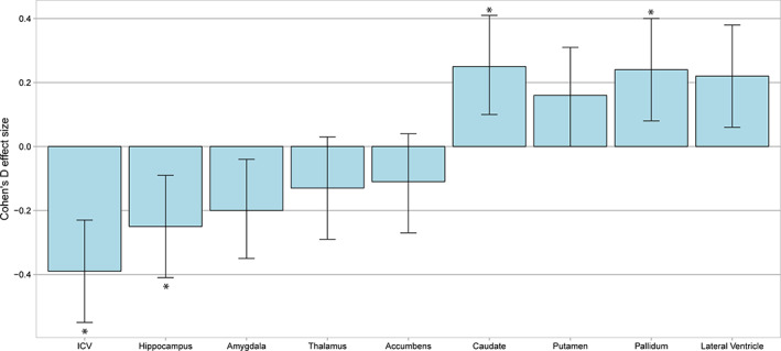
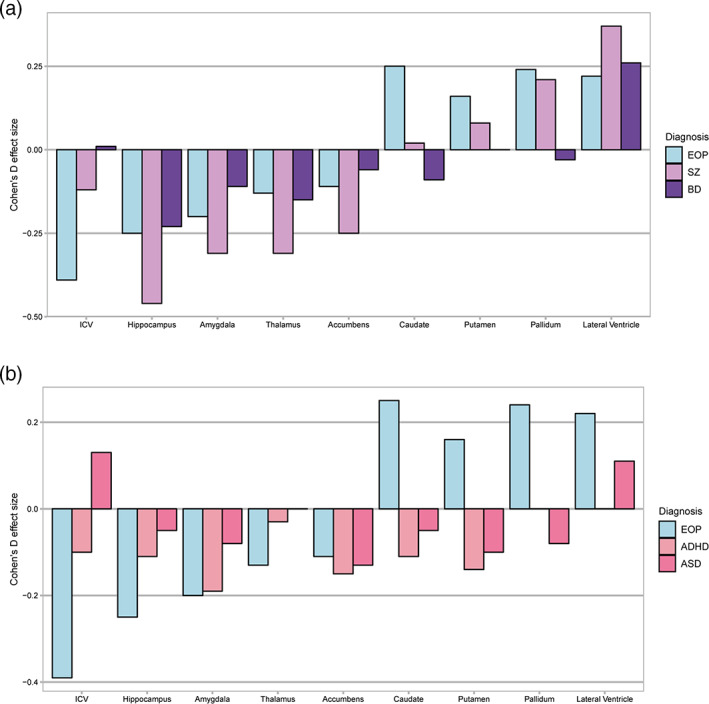
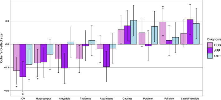
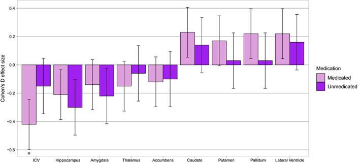
Early-onset psychosis disorders are serious mental disorders arising before the age of 18 years. Here, we investigate the largest neuroimaging dataset, to date, of patients with early-onset psychosis and healthy controls for differences in intracranial and subcortical brain volumes. The sample included 263 patients with early-onset psychosis (mean age: 16.4 ± 1.4 years, mean illness duration: 1.5 ± 1.4 years, 39.2% female) and 359 healthy controls (mean age: 15.9 ± 1.7 years, 45.4% female) with magnetic resonance imaging data, pooled from 11 clinical cohorts. Patients were diagnosed with early-onset schizophrenia (n = 183), affective psychosis (n = 39), or other psychotic disorders (n = 41). We used linear mixed-effects models to investigate differences in intracranial and subcortical volumes across the patient sample, diagnostic subgroup and antipsychotic medication, relative to controls. We observed significantly lower intracranial (Cohen's d = -0.39) and hippocampal (d = -0.25) volumes, and higher caudate (d = 0.25) and pallidum (d = 0.24) volumes in patients relative to controls. Intracranial volume was lower in both early-onset schizophrenia (d = -0.34) and affective psychosis (d = -0.42), and early-onset schizophrenia showed lower hippocampal (d = -0.24) and higher pallidum (d = 0.29) volumes. Patients who were currently treated with antipsychotic medication (n = 193) had significantly lower intracranial volume (d = -0.42). The findings demonstrate a similar pattern of brain alterations in early-onset psychosis as previously reported in adult psychosis, but with notably low intracranial volume. The low intracranial volume suggests disrupted neurodevelopment in adolescent early-onset psychosis.
早发性精神病障碍是指在 18 岁之前出现的严重精神障碍。在这里,我们研究了迄今为止最大的神经影像学数据集,以研究早发性精神病患者和健康对照者在颅内和皮质下脑容量方面的差异。该样本包括 263 名早发性精神病患者(平均年龄:16.4±1.4 岁,平均患病时间:1.5±1.4 年,39.2%为女性)和 359 名健康对照者(平均年龄:15.9±1.7 岁,45.4%为女性),他们的磁共振成像数据来自 11 个临床队列。患者被诊断为早发性精神分裂症(n=183)、情感性精神病(n=39)或其他精神病性障碍(n=41)。我们使用线性混合效应模型来研究患者样本、诊断亚组和抗精神病药物治疗之间的颅内和皮质下体积差异,与对照组相比。我们观察到患者的颅内(Cohen's d=-0.39)和海马(d=-0.25)体积明显降低,尾状核(d=0.25)和苍白球(d=0.24)体积明显升高。与对照组相比,早发性精神分裂症(d=-0.34)和情感性精神病(d=-0.42)患者的颅内体积均较低,早发性精神分裂症患者的海马体积较低(d=-0.24),苍白球体积较高(d=0.29)。目前正在接受抗精神病药物治疗的患者(n=193)颅内体积明显较低(d=-0.42)。研究结果表明,早发性精神病患者的脑结构改变与成人精神病患者先前报道的改变模式相似,但颅内体积明显较低。低颅内体积表明青少年早发性精神病的神经发育受到干扰。