The Maastricht MultiModal Molecular Imaging Institute (M4I), Maastricht University, Universiteitssingel 50, 6229 ER, Maastricht, The Netherlands.
Molecular Horizons and School of Chemistry and Molecular Bioscience, University of Wollongong, Wollongong, New South Wales, 2522, Australia.
Mol Imaging Biol. 2021 Apr;23(2):149-159. doi: 10.1007/s11307-020-01541-5. Epub 2020 Oct 6.
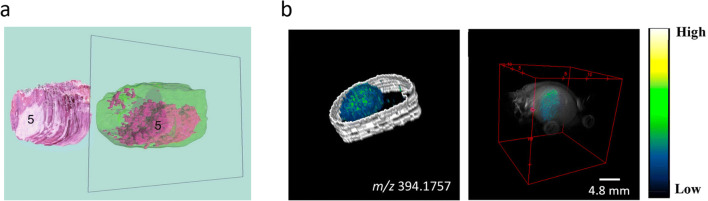
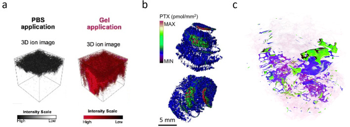
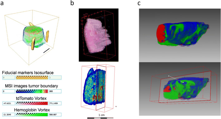
Mass spectrometry imaging (MSI) enables the visualization of molecular distributions on complex surfaces. It has been extensively used in the field of biomedical research to investigate healthy and diseased tissues. Most of the MSI studies are conducted in a 2D fashion where only a single slice of the full sample volume is investigated. However, biological processes occur within a tissue volume and would ideally be investigated as a whole to gain a more comprehensive understanding of the spatial and molecular complexity of biological samples such as tissues and cells. Mass spectrometry imaging has therefore been expanded to the 3D realm whereby molecular distributions within a 3D sample can be visualized. The benefit of investigating volumetric data has led to a quick rise in the application of single-sample 3D-MSI investigations. Several experimental and data analysis aspects need to be considered to perform successful 3D-MSI studies. In this review, we discuss these aspects as well as ongoing developments that enable 3D-MSI to be routinely applied to multi-sample studies.
质谱成像(MSI)能够在复杂表面上可视化分子分布。它已被广泛用于生物医学研究领域,以研究健康和患病组织。大多数 MSI 研究都是以 2D 方式进行的,其中仅研究整个样本体积的单个切片。然而,生物过程发生在组织体积内,理想情况下,应将其作为一个整体进行研究,以更全面地了解组织和细胞等生物样本的空间和分子复杂性。因此,质谱成像已扩展到 3D 领域,从而可以可视化 3D 样本内的分子分布。对体积数据进行研究的好处导致了单样本 3D-MSI 研究的快速应用。需要考虑几个实验和数据分析方面来成功进行 3D-MSI 研究。在这篇综述中,我们讨论了这些方面以及正在进行的开发,这些开发使 3D-MSI 能够常规地应用于多样本研究。