Škrátek Martin, Dvurečenskij Andrej, Kluknavský Michal, Barta Andrej, Bališ Peter, Mičurová Andrea, Cigáň Alexander, Eckstein-Andicsová Anita, Maňka Ján, Bernátová Iveta
Institute of Measurement Science, Slovak Academy of Sciences, 841 04 Bratislava, Slovakia.
Institute of Normal and Pathological Physiology, Centre of Experimental Medicine, Slovak Academy of Sciences, 813 71 Bratislava, Slovakia.
Nanomaterials (Basel). 2020 Oct 9;10(10):1993. doi: 10.3390/nano10101993.
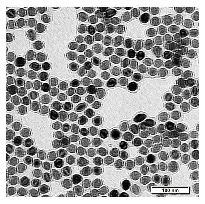
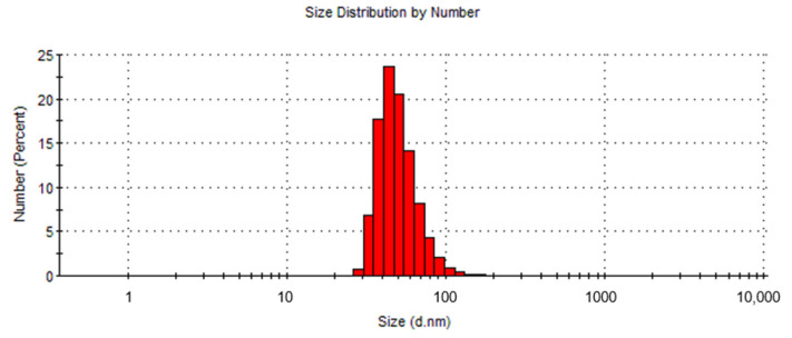
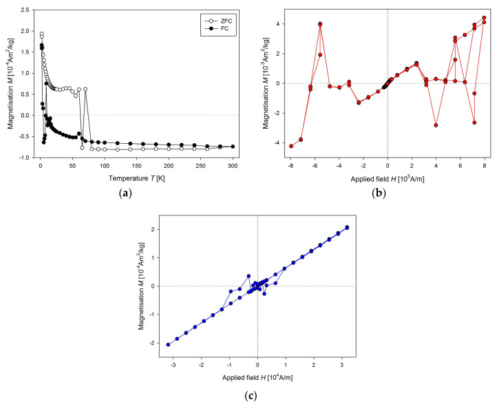
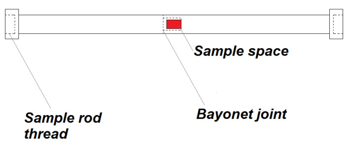
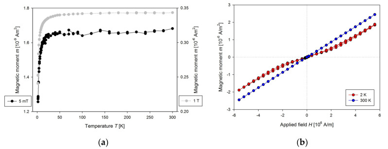
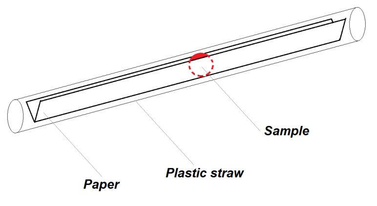
This study aimed to develop the method for determination of the ultra-small superparamagnetic iron oxide nanoparticle (USPION)-originated iron (UOI) in the tissues of rats on the basis of the magnetic characteristics (MC) in the liver, left heart ventricle (LHV), kidneys, aorta and blood of Wistar-Kyoto (WKY). Rats were treated intravenously by USPIONs dispersed in saline (transmission electron microscope (TEM) mean size ~30 nm, hydrodynamic size ~51 nm, nominal iron content 1 mg Fe/mL) at the low iron dose of 1 mg/kg. MC in the form of the mass magnetisation () versus the magnetic field () curves and temperature dependences of (determined using the SQUID magnetometer), histochemical determination of iron (by Perl's method) and USPION-induced superoxide production (by lucigenin-enhanced chemiluminescence) were investigated 100 min post-infusion. USPIONs significantly elevated superoxide production in the liver, LHV, kidney and aorta vs. the control group. Histochemical staining confirmed the presence of iron in all solid biological samples, however, this method was not suitable to unequivocally confirm the presence of UOI. We improved the SQUID magnetometric method and sample preparation to allow the determination of UOI by measurements of the MC of the tissues at 300 K in solid and liquid samples. The presence of the UOI was confirmed in all the tissues investigated in USPIONs-treated rats. The greatest levels were found in blood and lower amounts in the aorta, liver, LHV and kidneys. In conclusion, we have improved SQUID-magnetometric method to make it suitable for detection of low amounts of UOI in blood and tissues of rats.
本研究旨在基于Wistar-Kyoto(WKY)大鼠肝脏、左心室(LHV)、肾脏、主动脉和血液中的磁特性(MC),开发测定大鼠组织中超小超顺磁性氧化铁纳米颗粒(USPION)源性铁(UOI)的方法。大鼠通过静脉注射分散于生理盐水中的USPIONs(透射电子显微镜(TEM)平均尺寸约30nm,流体动力学尺寸约51nm,标称铁含量1mg Fe/mL),铁剂量为1mg/kg。在输注后100分钟,研究了质量磁化强度()与磁场()曲线形式的MC以及的温度依赖性(使用超导量子干涉仪磁强计测定)、铁的组织化学测定(采用Perl氏法)和USPION诱导的超氧化物生成(采用光泽精增强化学发光法)。与对照组相比,USPIONs显著提高了肝脏、LHV、肾脏和主动脉中的超氧化物生成。组织化学染色证实了所有固体生物样品中存在铁,然而,该方法不适用于明确确认UOI的存在。我们改进了超导量子干涉仪磁强计方法和样品制备,以便通过测量固体和液体样品在300K时组织的MC来测定UOI。在接受USPIONs处理的大鼠所研究的所有组织中均证实了UOI的存在。在血液中发现的含量最高,而在主动脉、肝脏、LHV和肾脏中的含量较低。总之,我们改进了超导量子干涉仪磁强计方法,使其适用于检测大鼠血液和组织中低含量的UOI。