Karolinska Institute, Institution for Clinical Sciences, Danderyd University Hospital, Stockholm, Sweden.
Acta Orthop. 2021 Feb;92(1):102-108. doi: 10.1080/17453674.2020.1837420. Epub 2020 Oct 26.
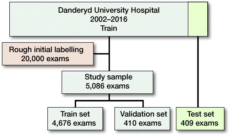
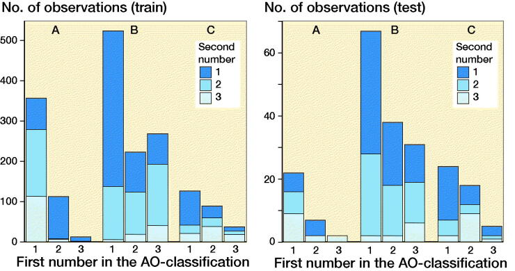
Background and purpose - Classification of ankle fractures is crucial for guiding treatment but advanced classifications such as the AO Foundation/Orthopedic Trauma Association (AO/OTA) are often too complex for human observers to learn and use. We have therefore investigated whether an automated algorithm that uses deep learning can learn to classify radiographs according to the new AO/OTA 2018 standards.Method - We trained a neural network based on the ResNet architecture on 4,941 radiographic ankle examinations. All images were classified according to the AO/OTA 2018 classification. A senior orthopedic surgeon (MG) then re-evaluated all images with fractures. We evaluated the network against a test set of 400 patients reviewed by 2 expert observers (MG, AS) independently.Results - In the training dataset, about half of the examinations contained fractures. The majority of the fractures were malleolar, of which the type B injuries represented almost 60% of the cases. Average area under the area under the receiver operating characteristic curve (AUC) was 0.90 (95% CI 0.82-0.94) for correctly classifying AO/OTA class where the most common major fractures, the malleolar type B fractures, reached an AUC of 0.93 (CI 0.90-0.95). The poorest performing type was malleolar A fractures, which included avulsions of the fibular tip.Interpretation - We found that a neural network could attain the required performance to aid with a detailed ankle fracture classification. This approach could be scaled up to other body parts. As the type of fracture is an important part of orthopedic decision-making, this is an important step toward computer-assisted decision-making.
背景与目的-踝关节骨折的分类对于指导治疗至关重要,但高级分类,如 AO 基金会/骨科创伤协会(AO/OTA)分类,对于人类观察者来说往往过于复杂,难以学习和使用。因此,我们研究了一种使用深度学习的自动算法是否可以学习根据新的 AO/OTA 2018 标准对 X 光片进行分类。
方法-我们基于 ResNet 架构训练了一个神经网络,使用了 4941 例放射学踝关节检查。所有图像均根据 AO/OTA 2018 分类进行分类。然后,一位资深骨科医生(MG)对所有骨折的图像进行重新评估。我们将该网络与由 2 位专家观察者(MG、AS)独立评估的 400 名患者的测试集进行了比较。
结果-在训练数据集中,大约一半的检查包含骨折。大多数骨折为外踝骨折,其中 B 型损伤占近 60%的病例。正确分类 AO/OTA 类别的平均受试者工作特征曲线下面积(AUC)为 0.90(95%CI 0.82-0.94),其中最常见的主要骨折,即外踝 B 型骨折,AUC 达到 0.93(CI 0.90-0.95)。表现最差的类型是外踝 A 型骨折,其中包括腓骨尖端的撕脱。
解释-我们发现神经网络可以达到辅助详细踝关节骨折分类所需的性能。这种方法可以扩展到其他身体部位。由于骨折类型是骨科决策的重要组成部分,这是迈向计算机辅助决策的重要一步。