Kammoun Rym, Zmantar Tarek, Ghoul Sonia
Laboratory of Histology and Embryology, Faculty of Dental Medicine, University of Monastir, Tunisia.
Laboratory of Dento-Facial, Clinical and Biological Approach (ABCDF), Tunisia.
MethodsX. 2020 Oct 17;7:101107. doi: 10.1016/j.mex.2020.101107. eCollection 2020.
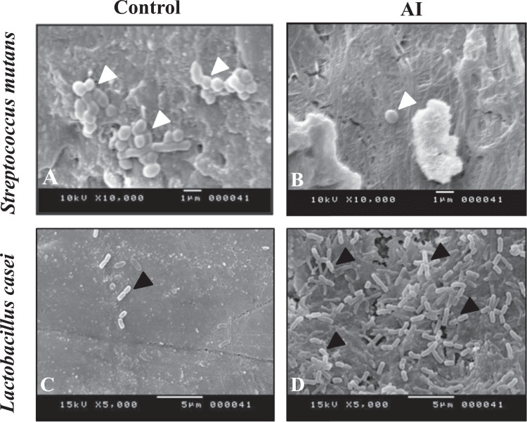
To describe the bacterial adhesion protocol of and asei on dental surfaces for a qualitative approach by Scanning Electron Microscopy (SEM) observations. A control and Amelogenesis Imperfecta (AI) affected teeth were used to validate the protocol.
Eight teeth were collected and fixed in 10% formalin during 10 days. Crowns were fragmented into 4 parts and kept in the freshly prepared artificial saliva. For the preparation of bacterial suspensions, bacterial strains ( and L. ) were incubated in a freshly prepared culture medium. After two successive cultures at 37 °C and 3 rinces, bacterial suspensions were prepared in artificial saliva and adjusted to correspond to 10 CFU ml. Bacterial adhesion was carried out by sedimentation. Dental fragments were immersed in bacterial suspensions and rinsed with PBS to remove non adherent bacteria. Adherent bacteria were fixed with glutaraldehyde. Finally, teeth samples were dehydrated, coated, dried and observed using high-resolution SEM (JEOL, JSM-5400).
SEM observations showed adherence of spheric stuctures, identified as and bacilic structures identified as L. .
Adhesion of bacteria could be observed by SEM and depends on the quality of dental mineralized tissues.
通过扫描电子显微镜(SEM)观察,描述用于定性研究的变形链球菌和远缘链球菌在牙面的细菌黏附方案。使用对照牙和患有牙釉质发育不全(AI)的牙齿来验证该方案。
收集八颗牙齿并在10%福尔马林中固定10天。将牙冠切成4部分并保存在新制备的人工唾液中。为制备细菌悬液,将细菌菌株(变形链球菌和远缘链球菌)在新制备的培养基中培养。在37℃连续培养两次并冲洗3次后,在人工唾液中制备细菌悬液并调整至对应10⁸CFU/ml。通过沉降法进行细菌黏附。将牙齿碎片浸入细菌悬液中,并用磷酸盐缓冲液(PBS)冲洗以去除未黏附的细菌。用戊二醛固定黏附的细菌。最后,对牙齿样本进行脱水、镀膜、干燥,并使用高分辨率扫描电子显微镜(JEOL,JSM - 5400)进行观察。
扫描电子显微镜观察显示有球形结构(鉴定为变形链球菌)和杆状结构(鉴定为远缘链球菌)的黏附。
通过扫描电子显微镜可以观察到细菌的黏附,且其取决于牙矿化组织的质量。