• 文献检索
  • 文档翻译
  • 深度研究
  • 学术资讯
  • Suppr Zotero 插件Zotero 插件
  • 邀请有礼
  • 套餐&价格
  • 历史记录
应用&插件
Suppr Zotero 插件Zotero 插件浏览器插件Mac 客户端Windows 客户端微信小程序
定价
高级版会员购买积分包购买API积分包
服务
文献检索文档翻译深度研究API 文档MCP 服务
关于我们
关于 Suppr公司介绍联系我们用户协议隐私条款
关注我们

Suppr 超能文献

核心技术专利:CN118964589B侵权必究
粤ICP备2023148730 号-1Suppr @ 2026

文献检索

告别复杂PubMed语法,用中文像聊天一样搜索,搜遍4000万医学文献。AI智能推荐,让科研检索更轻松。

立即免费搜索

文件翻译

保留排版,准确专业,支持PDF/Word/PPT等文件格式,支持 12+语言互译。

免费翻译文档

深度研究

AI帮你快速写综述,25分钟生成高质量综述,智能提取关键信息,辅助科研写作。

立即免费体验

哺乳动物细胞的 3D 微组织打印以生成生物组织。

3D micro-organisation printing of mammalian cells to generate biological tissues.

机构信息

Fluicell AB, Flöjelbergsgatan 8C, 431 37, Mölndal, Sweden.

Cellectricon AB, Neongatan 4B, 431 53, Mölndal, Sweden.

出版信息

Sci Rep. 2020 Nov 10;10(1):19529. doi: 10.1038/s41598-020-74191-w.

DOI:10.1038/s41598-020-74191-w
PMID:33173097
原文链接:https://pmc.ncbi.nlm.nih.gov/articles/PMC7656262/
Abstract

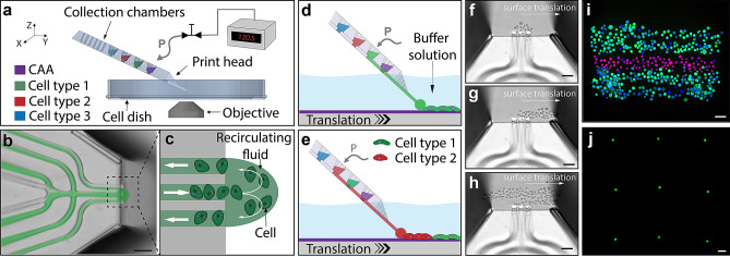
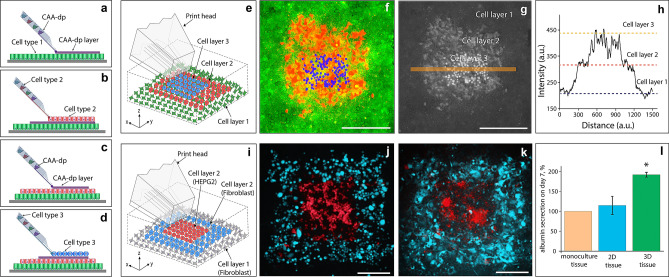
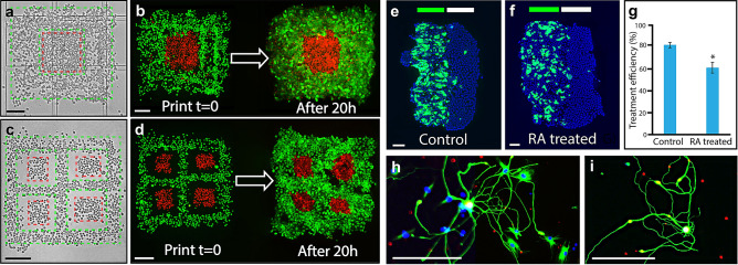
Significant strides have been made in the development of in vitro systems for disease modelling. However, the requirement of microenvironment control has placed limitations on the generation of relevant models. Herein, we present a biological tissue printing approach that employs open-volume microfluidics to position individual cells in complex 2D and 3D patterns, as well as in single cell arrays. The variety of bioprinted cell types employed, including skin epithelial (HaCaT), skin cancer (A431), liver cancer (Hep G2), and fibroblast (3T3-J2) cells, all of which exhibited excellent viability and survivability, allowing printed structures to rapidly develop into confluent tissues. To demonstrate a simple 2D oncology model, A431 and HaCaT cells were printed and grown into tissues. Furthermore, a basic skin model was established to probe drug response. 3D tissue formation was demonstrated by co-printing Hep G2 and 3T3-J2 cells onto an established fibroblast layer, the functionality of which was probed by measuring albumin production, and was found to be higher in comparison to both 2D and monoculture approaches. Bioprinting of primary cells was tested using acutely isolated primary rat dorsal root ganglia neurons, which survived and established processes. The presented technique offers a novel open-volume microfluidics approach to bioprint cells for the generation of biological tissues.

摘要

在疾病建模的体外系统开发方面已经取得了重大进展。然而,微环境控制的要求对相关模型的生成提出了限制。在此,我们提出了一种生物组织打印方法,该方法采用开体积微流控技术将单个细胞定位在复杂的 2D 和 3D 图案中,以及单细胞阵列中。所使用的各种生物打印细胞类型,包括皮肤上皮细胞(HaCaT)、皮肤癌细胞(A431)、肝癌细胞(Hep G2)和成纤维细胞(3T3-J2),所有这些细胞都表现出出色的活力和生存能力,使打印结构能够迅速发展为融合组织。为了展示一个简单的 2D 肿瘤模型,我们打印了 A431 和 HaCaT 细胞并使其生长为组织。此外,还建立了一个基本的皮肤模型来探测药物反应。通过将 Hep G2 和 3T3-J2 细胞共打印到已建立的成纤维细胞层上,实现了 3D 组织的形成,通过测量白蛋白的产生来探测其功能,发现与 2D 和单培养方法相比,其功能更高。使用急性分离的大鼠背根神经节神经元测试了原代细胞的生物打印,这些神经元存活并建立了过程。所提出的技术为生物打印细胞生成生物组织提供了一种新颖的开体积微流控方法。

https://cdn.ncbi.nlm.nih.gov/pmc/blobs/049b/7656262/0e045efa3819/41598_2020_74191_Fig3_HTML.jpg
https://cdn.ncbi.nlm.nih.gov/pmc/blobs/049b/7656262/6e179bd6d35b/41598_2020_74191_Fig1_HTML.jpg
https://cdn.ncbi.nlm.nih.gov/pmc/blobs/049b/7656262/ee46d937073b/41598_2020_74191_Fig2_HTML.jpg
https://cdn.ncbi.nlm.nih.gov/pmc/blobs/049b/7656262/0e045efa3819/41598_2020_74191_Fig3_HTML.jpg
https://cdn.ncbi.nlm.nih.gov/pmc/blobs/049b/7656262/6e179bd6d35b/41598_2020_74191_Fig1_HTML.jpg
https://cdn.ncbi.nlm.nih.gov/pmc/blobs/049b/7656262/ee46d937073b/41598_2020_74191_Fig2_HTML.jpg
https://cdn.ncbi.nlm.nih.gov/pmc/blobs/049b/7656262/0e045efa3819/41598_2020_74191_Fig3_HTML.jpg

相似文献

1
3D micro-organisation printing of mammalian cells to generate biological tissues.哺乳动物细胞的 3D 微组织打印以生成生物组织。
Sci Rep. 2020 Nov 10;10(1):19529. doi: 10.1038/s41598-020-74191-w.
2
Human stem cell based corneal tissue mimicking structures using laser-assisted 3D bioprinting and functional bioinks.利用激光辅助 3D 生物打印和功能生物墨水构建基于人干细胞的角膜组织模拟结构。
Biomaterials. 2018 Jul;171:57-71. doi: 10.1016/j.biomaterials.2018.04.034. Epub 2018 Apr 16.
3
3D-bioprinted all-inclusive bioanalytical platforms for cell studies.3D 生物打印全包容型生物分析平台用于细胞研究。
Sci Rep. 2020 Sep 4;10(1):14669. doi: 10.1038/s41598-020-71452-6.
4
Three-dimensional direct cell bioprinting for tissue engineering.三维直接细胞生物打印在组织工程中的应用。
J Biomed Mater Res B Appl Biomater. 2017 Nov;105(8):2530-2544. doi: 10.1002/jbm.b.33768. Epub 2016 Sep 30.
5
Bioprinting of human pluripotent stem cells and their directed differentiation into hepatocyte-like cells for the generation of mini-livers in 3D.人类多能干细胞的生物打印及其定向分化为类肝细胞以生成三维微型肝脏。
Biofabrication. 2015 Oct 21;7(4):044102. doi: 10.1088/1758-5090/7/4/044102.
6
Functional characterization of 3D contractile smooth muscle tissues generated using a unique microfluidic 3D bioprinting technology.使用独特的微流控 3D 生物打印技术生成的 3D 收缩性平滑肌组织的功能表征。
FASEB J. 2020 Jan;34(1):1652-1664. doi: 10.1096/fj.201901063RR. Epub 2019 Dec 4.
7
Proof-of-concept: 3D bioprinting of pigmented human skin constructs.概念验证:彩色人种皮肤结构的 3D 生物打印。
Biofabrication. 2018 Jan 23;10(2):025005. doi: 10.1088/1758-5090/aa9e1e.
8
Microfluidic bioprinting for organ-on-a-chip models.微流控生物打印在器官芯片模型中的应用。
Drug Discov Today. 2019 Jun;24(6):1248-1257. doi: 10.1016/j.drudis.2019.03.025. Epub 2019 Mar 30.
9
Feasibility of Bioprinting with a Modified Desktop 3D Printer.使用改良桌面3D打印机进行生物打印的可行性
Tissue Eng Part C Methods. 2016 Dec;22(12):1071-1076. doi: 10.1089/ten.TEC.2016.0286.
10
3D modeling of normal skin and cutaneous squamous cell carcinoma. A comparative study in 2D cultures, spheroids, and 3D bioprinted systems.正常皮肤和皮肤鳞状细胞癌的3D建模。在2D培养、球体和3D生物打印系统中的比较研究。
Biofabrication. 2024 Feb 29;16(2). doi: 10.1088/1758-5090/ad2b06.

引用本文的文献

1
Bio-printing method as a novel approach to obtain a fibrin scaffold settled by limbal epithelial cells for corneal regeneration.生物打印方法作为一种获得纤维蛋白支架的新方法,该支架中定居有角膜缘上皮细胞,可用于角膜再生。
Sci Rep. 2024 Oct 7;14(1):23352. doi: 10.1038/s41598-024-73383-y.
2
Systematic Review of Genetic Substrate Reduction Therapy in Lysosomal Storage Diseases: Opportunities, Challenges and Delivery Systems.系统性综述溶酶体贮积症中基因底物减少疗法:机遇、挑战与传递系统。
BioDrugs. 2024 Sep;38(5):657-680. doi: 10.1007/s40259-024-00674-1. Epub 2024 Aug 23.
3
3D bioprinting of liver models: A systematic scoping review of methods, bioinks, and reporting quality.

本文引用的文献

1
The bioprinting roadmap.生物打印路线图。
Biofabrication. 2020 Feb 6;12(2):022002. doi: 10.1088/1758-5090/ab5158.
2
Progress in 3D bioprinting technology for tissue/organ regenerative engineering.用于组织/器官再生工程的3D生物打印技术进展。
Biomaterials. 2020 Jan;226:119536. doi: 10.1016/j.biomaterials.2019.119536. Epub 2019 Oct 11.
3
3D bioprinting of collagen to rebuild components of the human heart.3D 生物打印胶原蛋白以重建人类心脏的组件。
肝脏模型的3D生物打印:方法、生物墨水及报告质量的系统综述
Mater Today Bio. 2024 Feb 15;26:100991. doi: 10.1016/j.mtbio.2024.100991. eCollection 2024 Jun.
4
Multiplex Single-Cell Bioprinting for Engineering of Heterogeneous Tissue Constructs with Subcellular Spatial Resolution.用于构建具有亚细胞空间分辨率的异质组织构建体的多重单细胞生物打印
bioRxiv. 2024 Feb 11:2024.02.01.578499. doi: 10.1101/2024.02.01.578499.
5
Advancements of 3D bioprinting in regenerative medicine: Exploring cell sources for organ fabrication.3D生物打印在再生医学中的进展:探索用于器官制造的细胞来源。
Heliyon. 2024 Jan 17;10(3):e24593. doi: 10.1016/j.heliyon.2024.e24593. eCollection 2024 Feb 15.
6
Application of three-dimensional (3D) bioprinting in anti-cancer therapy.三维(3D)生物打印在抗癌治疗中的应用。
Heliyon. 2023 Sep 28;9(10):e20475. doi: 10.1016/j.heliyon.2023.e20475. eCollection 2023 Oct.
7
Opportunities and challenges to engineer 3D models of tumor-adaptive immune interactions.构建肿瘤适应性免疫相互作用的 3D 模型的机遇与挑战。
Front Immunol. 2023 Apr 4;14:1162905. doi: 10.3389/fimmu.2023.1162905. eCollection 2023.
8
Hydrogels for Tissue Engineering: Addressing Key Design Needs Toward Clinical Translation.用于组织工程的水凝胶:满足临床转化的关键设计需求
Front Bioeng Biotechnol. 2022 May 5;10:849831. doi: 10.3389/fbioe.2022.849831. eCollection 2022.
9
Bioprinting of Complex Multicellular Organs with Advanced Functionality-Recent Progress and Challenges Ahead.具有先进功能的复杂多细胞器官的生物打印-最新进展和未来挑战。
Adv Mater. 2022 Jan;34(3):e2101321. doi: 10.1002/adma.202101321. Epub 2021 Nov 5.
10
Facile Cell-Friendly Hollow-Core Fiber Diffusion-Limited Photofabrication.简易的细胞友好型中空纤维扩散限制光制造
Front Bioeng Biotechnol. 2021 Dec 3;9:783834. doi: 10.3389/fbioe.2021.783834. eCollection 2021.
Science. 2019 Aug 2;365(6452):482-487. doi: 10.1126/science.aav9051.
4
Effective bioprinting resolution in tissue model fabrication.组织模型制造中的有效生物打印分辨率。
Lab Chip. 2019 Jun 7;19(11):2019-2037. doi: 10.1039/c8lc01037d. Epub 2019 May 13.
5
Microfluidic bioprinting for organ-on-a-chip models.微流控生物打印在器官芯片模型中的应用。
Drug Discov Today. 2019 Jun;24(6):1248-1257. doi: 10.1016/j.drudis.2019.03.025. Epub 2019 Mar 30.
6
Clinical-grade stem cell-derived retinal pigment epithelium patch rescues retinal degeneration in rodents and pigs.临床级别的干细胞衍生的视网膜色素上皮贴片可挽救啮齿动物和猪的视网膜变性。
Sci Transl Med. 2019 Jan 16;11(475). doi: 10.1126/scitranslmed.aat5580.
7
3D Inkjet Printing of Complex, Cell-Laden Hydrogel Structures.3D 喷墨打印复杂的、含细胞的水凝胶结构。
Sci Rep. 2018 Nov 20;8(1):17099. doi: 10.1038/s41598-018-35504-2.
8
Network-Based Drug Discovery: Coupling Network Pharmacology with Phenotypic Screening for Neuronal Excitability.基于网络的药物发现:将网络药理学与神经元兴奋性表型筛选相结合。
J Mol Biol. 2018 Sep 14;430(18 Pt A):3005-3015. doi: 10.1016/j.jmb.2018.07.016. Epub 2018 Jul 18.
9
Three-dimensional bioprinting of stem-cell derived tissues for human regenerative medicine.基于干细胞的组织的三维生物打印用于人类再生医学。
Philos Trans R Soc Lond B Biol Sci. 2018 Jul 5;373(1750). doi: 10.1098/rstb.2017.0224.
10
Phase 1 clinical study of an embryonic stem cell-derived retinal pigment epithelium patch in age-related macular degeneration.胚胎干细胞源性视网膜色素上皮贴片中年龄相关性黄斑变性的 1 期临床研究。
Nat Biotechnol. 2018 Apr;36(4):328-337. doi: 10.1038/nbt.4114. Epub 2018 Mar 19.