Department of Ophthalmology, School of Medicine, Gunma University, 3-39-15 Showa-machi, Maebashi, Gunma, 371-8511, Japan.
Sci Rep. 2020 Nov 11;10(1):19505. doi: 10.1038/s41598-020-75789-w.
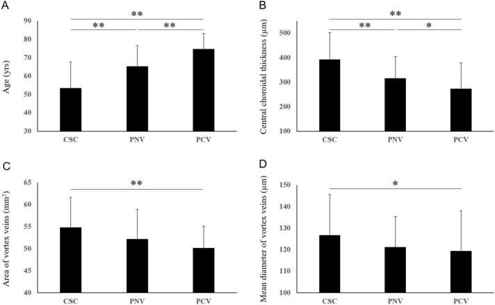
Pachychoroid spectrum diseases have attracted increasing attention, though their pathophysiology has yet to be fully elucidated. In this study, we assessed the vascular diameters of vortex veins in pachychoroid spectrum diseases such as central serous chorioretinopathy (CSC), pachychoroid neovasculopathy without polypoidal lesions (PNV), and pachychoroid neovasculopathy with polypoidal lesions (polypoidal choroidal vasculopathy: PCV). In a retrospective case series of 94 eyes with CSC, 60 eyes with PNV and 57 with PCV, we binarized en face optical coherence tomography (OCT) images of choroidal vortex veins and analyzed the mean diameter of vortex veins. The presence of anastomosis between the superior and inferior vortex veins and central choroidal thickness (CCT) were also evaluated using OCT images. CSC showed significantly larger mean diameter of vortex veins than PCV (P < 0.05). Anastomosis between superior and inferior vortex veins was observed in over 90% of eyes with each pachychoroid spectrum disease. The patients with CSC were the youngest, followed by PNV patients, and then patients with PCV. The largest CCT values were observed in CSC eyes, followed by PNV eyes, and then PCV eyes. CCT correlated with the mean diameter of vortex veins (rs = 0.51, P < 0.01). These findings suggest that congestion of vortex veins might show gradual amelioration corresponding to the development of anastomosis between the superior and inferior vortex veins during the course of progression of pachychoroid spectrum diseases. Moreover, the mean diameter of vortex veins can be used as a parameter indicating choroidal congestion.
脉络膜增厚谱疾病越来越受到关注,但其病理生理学尚未完全阐明。在这项研究中,我们评估了中心性浆液性脉络膜视网膜病变(CSC)、无息肉状脉络膜血管病变(PNV)和有息肉状脉络膜血管病变(息肉状脉络膜血管病变:PCV)等脉络膜增厚谱疾病的涡静脉血管直径。在一项回顾性病例系列研究中,纳入了 94 只 CSC 眼、60 只 PNV 眼和 57 只 PCV 眼,我们对脉络膜涡静脉的面内光学相干断层扫描(OCT)图像进行了二值化处理,并分析了涡静脉的平均直径。还使用 OCT 图像评估了上下涡静脉之间的吻合和脉络膜中央厚度(CCT)。CSC 的涡静脉平均直径明显大于 PCV(P<0.05)。每种脉络膜增厚谱疾病的眼都观察到超过 90%的上下涡静脉吻合。CSC 患者最年轻,其次是 PNV 患者,然后是 PCV 患者。CCT 值最大的是 CSC 眼,其次是 PNV 眼,然后是 PCV 眼。CCT 与涡静脉平均直径呈正相关(rs=0.51,P<0.01)。这些发现表明,在脉络膜增厚谱疾病进展过程中,随着上下涡静脉之间吻合的发展,涡静脉充血可能逐渐缓解。此外,涡静脉的平均直径可用作指示脉络膜充血的参数。