Department of Materials, Department of Bioengineering and Institute of Biomedical Engineering, Imperial College London, London, SW7 2AZ, UK.
Department of Craniofacial Development & Stem Cell Biology, Kings College London, Tower Wing, Guy's Hospital, London, SE1 9RT, UK.
Nat Commun. 2020 Dec 2;11(1):6172. doi: 10.1038/s41467-020-19827-1.
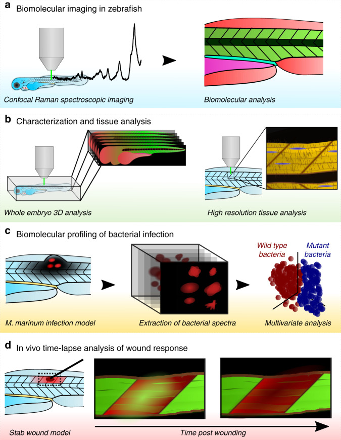
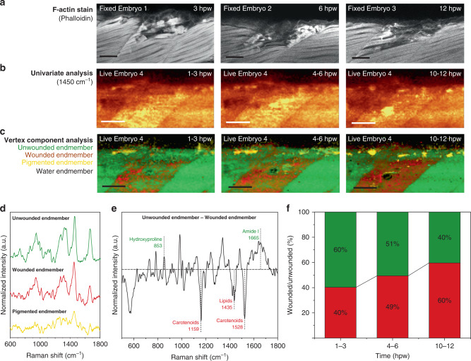
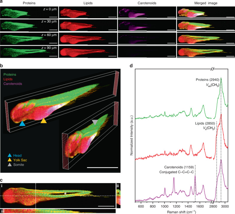
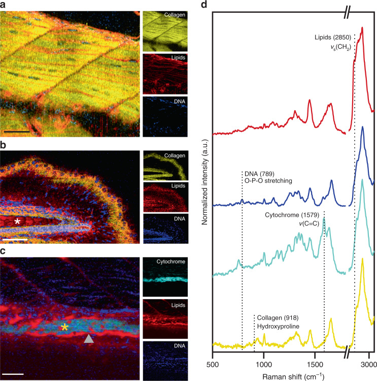
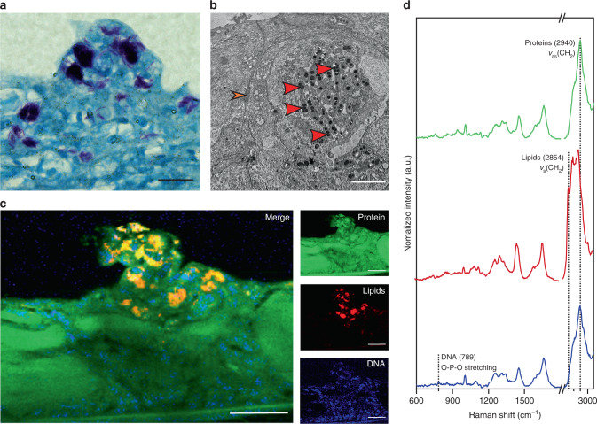
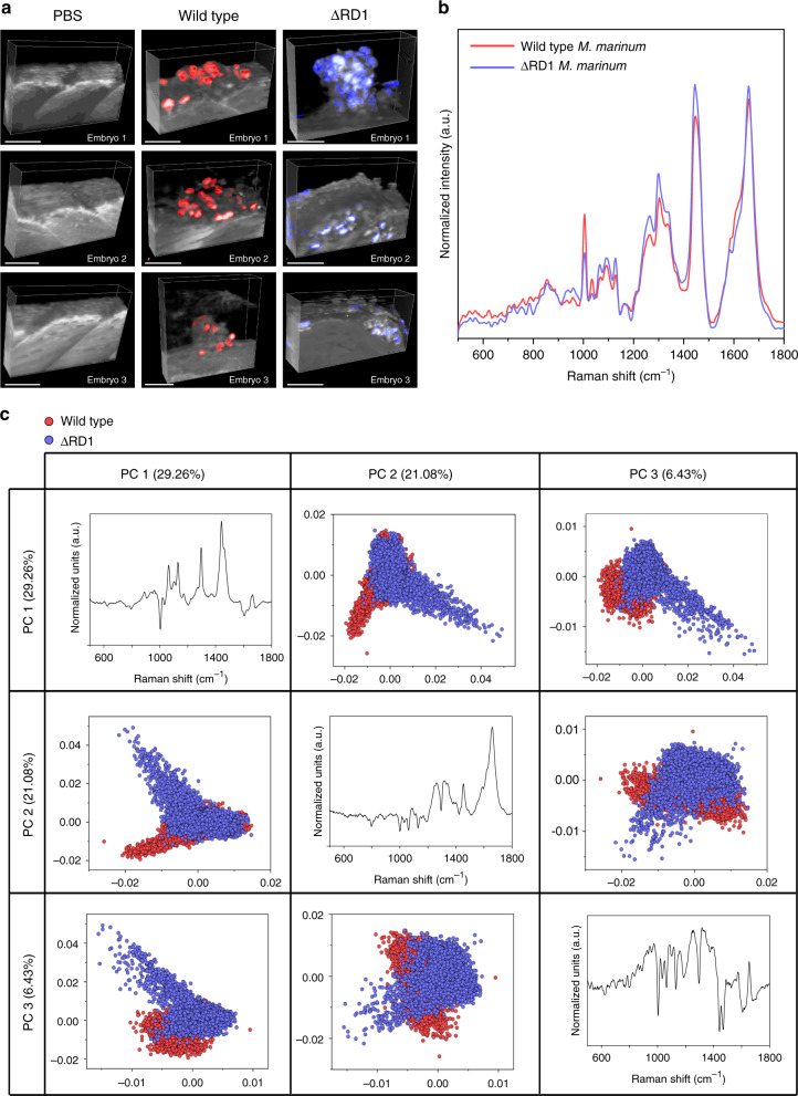
Zebrafish embryos provide a unique opportunity to visualize complex biological processes, yet conventional imaging modalities are unable to access intricate biomolecular information without compromising the integrity of the embryos. Here, we report the use of confocal Raman spectroscopic imaging for the visualization and multivariate analysis of biomolecular information extracted from unlabeled zebrafish embryos. We outline broad applications of this method in: (i) visualizing the biomolecular distribution of whole embryos in three dimensions, (ii) resolving anatomical features at subcellular spatial resolution, (iii) biomolecular profiling and discrimination of wild type and ΔRD1 mutant Mycobacterium marinum strains in a zebrafish embryo model of tuberculosis and (iv) in vivo temporal monitoring of the wound response in living zebrafish embryos. Overall, this study demonstrates the application of confocal Raman spectroscopic imaging for the comparative bimolecular analysis of fully intact and living zebrafish embryos.
斑马鱼胚胎为可视化复杂的生物过程提供了独特的机会,但传统的成像方式在不破坏胚胎完整性的情况下,无法获取复杂的生物分子信息。在这里,我们报告了使用共焦拉曼光谱成像技术对未标记的斑马鱼胚胎中提取的生物分子信息进行可视化和多元分析。我们概述了该方法在以下方面的广泛应用:(i)在三维空间中可视化整个胚胎的生物分子分布,(ii)以亚细胞空间分辨率解析解剖特征,(iii)在结核分枝杆菌斑马鱼胚胎模型中对野生型和ΔRD1 突变株进行生物分子分析和区分,以及(iv)在活斑马鱼胚胎中进行体内伤口反应的时间监测。总的来说,这项研究证明了共焦拉曼光谱成像在比较完整和活体斑马鱼胚胎的比较生物分子分析中的应用。