文献检索文档翻译深度研究
Suppr Zotero 插件Zotero 插件
邀请有礼套餐&价格历史记录

新学期,新优惠

限时优惠:9月1日-9月22日

30天高级会员仅需29元

1天体验卡首发特惠仅需5.99元

了解详情
不再提醒
插件&应用
Suppr Zotero 插件Zotero 插件浏览器插件Mac 客户端Windows 客户端微信小程序
高级版
套餐订阅购买积分包
AI 工具
文献检索文档翻译深度研究
关于我们
关于 Suppr公司介绍联系我们用户协议隐私条款
关注我们

Suppr 超能文献

核心技术专利:CN118964589B侵权必究
粤ICP备2023148730 号-1Suppr @ 2025

优化遗传算法-极限学习机方法自动检测 COVID-19。

Optimised genetic algorithm-extreme learning machine approach for automatic COVID-19 detection.

机构信息

CAIT, Faculty of Information Science and Technology, Universiti Kebangsaan Malaysia, Bangi, Selangor, Malaysia.

Department of Communication Engineering, School of Electrical Engineering, Universiti Teknologi Malaysia, UTM Johor Bahru, Johor, Malaysia.

出版信息

PLoS One. 2020 Dec 15;15(12):e0242899. doi: 10.1371/journal.pone.0242899. eCollection 2020.


DOI:10.1371/journal.pone.0242899
PMID:33320858
原文链接:https://pmc.ncbi.nlm.nih.gov/articles/PMC7737907/
Abstract

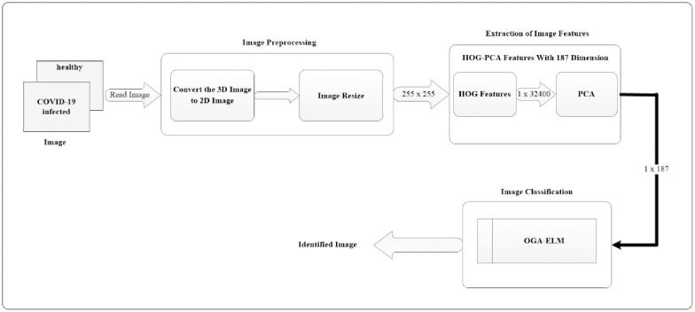
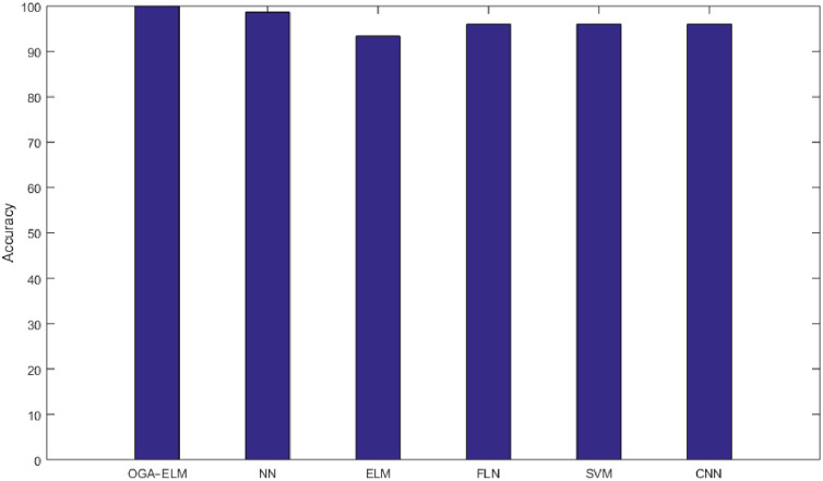
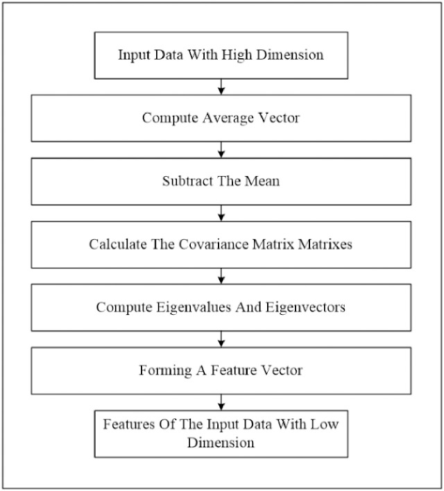
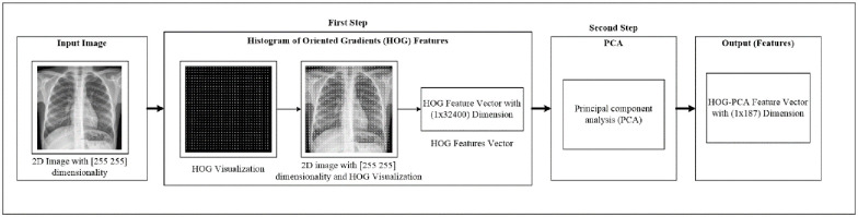
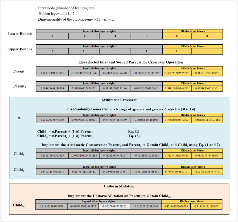
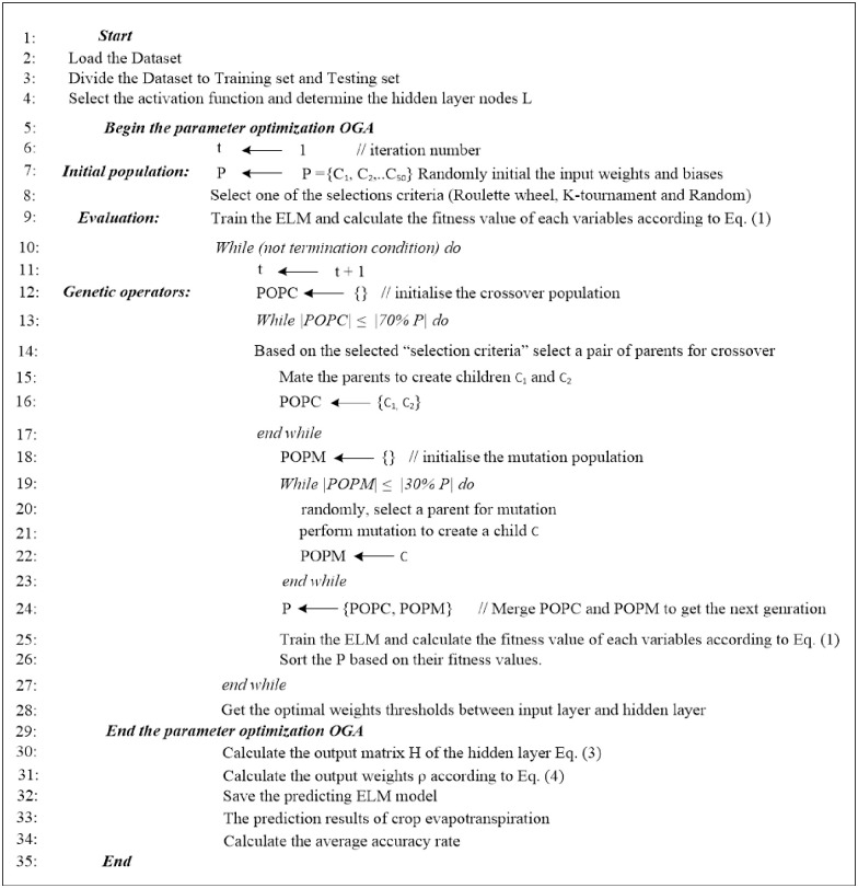
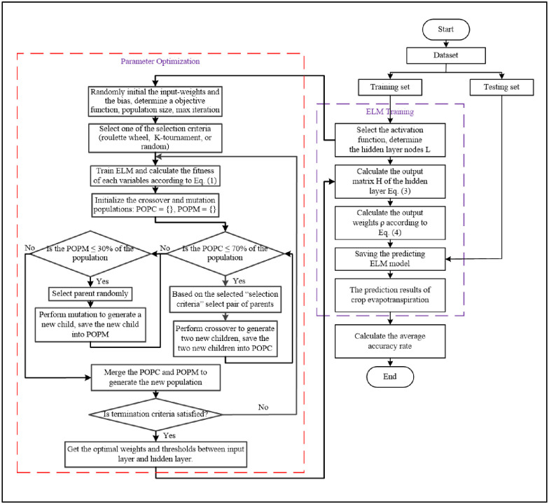
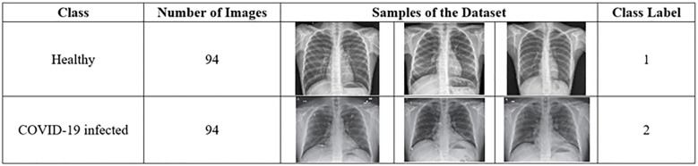
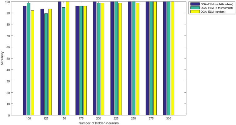
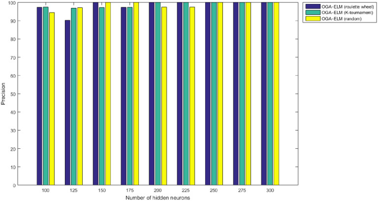
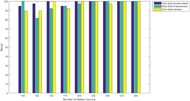
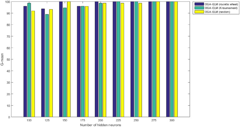
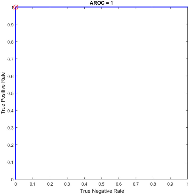
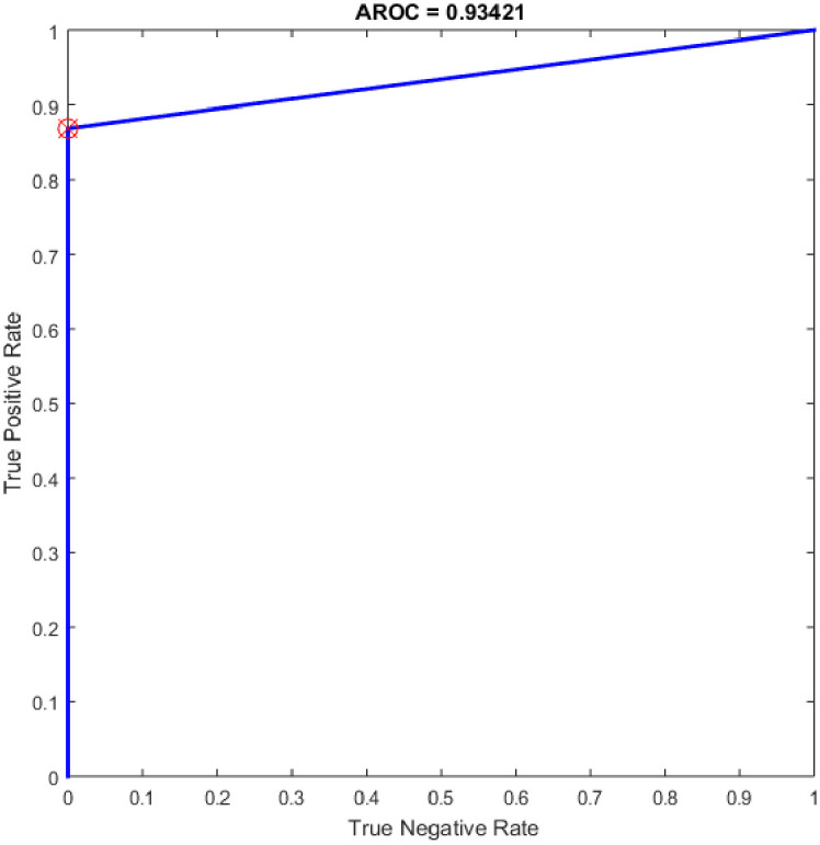
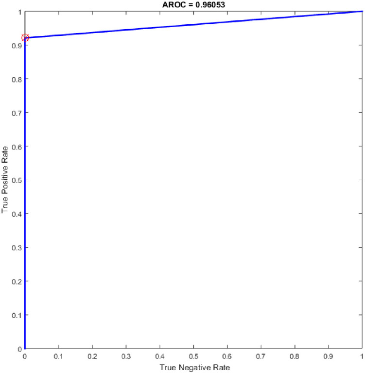
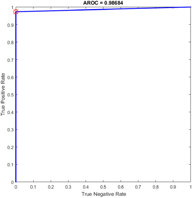
The coronavirus disease (COVID-19), is an ongoing global pandemic caused by severe acute respiratory syndrome. Chest Computed Tomography (CT) is an effective method for detecting lung illnesses, including COVID-19. However, the CT scan is expensive and time-consuming. Therefore, this work focus on detecting COVID-19 using chest X-ray images because it is widely available, faster, and cheaper than CT scan. Many machine learning approaches such as Deep Learning, Neural Network, and Support Vector Machine; have used X-ray for detecting the COVID-19. Although the performance of those approaches is acceptable in terms of accuracy, however, they require high computational time and more memory space. Therefore, this work employs an Optimised Genetic Algorithm-Extreme Learning Machine (OGA-ELM) with three selection criteria (i.e., random, K-tournament, and roulette wheel) to detect COVID-19 using X-ray images. The most crucial strength factors of the Extreme Learning Machine (ELM) are: (i) high capability of the ELM in avoiding overfitting; (ii) its usability on binary and multi-type classifiers; and (iii) ELM could work as a kernel-based support vector machine with a structure of a neural network. These advantages make the ELM efficient in achieving an excellent learning performance. ELMs have successfully been applied in many domains, including medical domains such as breast cancer detection, pathological brain detection, and ductal carcinoma in situ detection, but not yet tested on detecting COVID-19. Hence, this work aims to identify the effectiveness of employing OGA-ELM in detecting COVID-19 using chest X-ray images. In order to reduce the dimensionality of a histogram oriented gradient features, we use principal component analysis. The performance of OGA-ELM is evaluated on a benchmark dataset containing 188 chest X-ray images with two classes: a healthy and a COVID-19 infected. The experimental result shows that the OGA-ELM achieves 100.00% accuracy with fast computation time. This demonstrates that OGA-ELM is an efficient method for COVID-19 detecting using chest X-ray images.

摘要

冠状病毒病(COVID-19)是一种由严重急性呼吸系统综合征引起的持续全球大流行疾病。胸部计算机断层扫描(CT)是一种有效的检测肺部疾病的方法,包括 COVID-19。然而,CT 扫描昂贵且耗时。因此,这项工作专注于使用胸部 X 射线图像检测 COVID-19,因为它广泛可用,比 CT 扫描更快且更便宜。许多机器学习方法,如深度学习、神经网络和支持向量机;已经使用 X 射线来检测 COVID-19。尽管这些方法在准确性方面的性能可以接受,但是它们需要高计算时间和更多的内存空间。因此,这项工作采用了三种选择标准(即随机、K-锦标赛和轮盘赌)的优化遗传算法-极限学习机(OGA-ELM)来使用 X 射线图像检测 COVID-19。极限学习机(ELM)的最重要的关键优势是:(i)ELM 在避免过拟合方面的能力很高;(ii)它在二进制和多类分类器中的可用性;以及(iii)ELM 可以作为具有神经网络结构的核支持向量机工作。这些优势使 ELM 在实现出色的学习性能方面非常高效。ELM 已经成功应用于许多领域,包括乳腺癌检测、病理性脑检测和原位导管癌检测等医学领域,但尚未在 COVID-19 检测中进行测试。因此,这项工作旨在确定使用胸部 X 射线图像使用 OGA-ELM 检测 COVID-19 的有效性。为了降低直方图定向梯度特征的维数,我们使用主成分分析。OGA-ELM 的性能在包含 188 张胸部 X 射线图像的基准数据集上进行评估,这些图像分为两类:健康和 COVID-19 感染。实验结果表明,OGA-ELM 以快速的计算时间实现了 100.00%的准确率。这表明 OGA-ELM 是一种使用胸部 X 射线图像检测 COVID-19 的有效方法。

https://cdn.ncbi.nlm.nih.gov/pmc/blobs/705a/7737907/1b66d7581878/pone.0242899.g023.jpg
https://cdn.ncbi.nlm.nih.gov/pmc/blobs/705a/7737907/84943135cc91/pone.0242899.g001.jpg
https://cdn.ncbi.nlm.nih.gov/pmc/blobs/705a/7737907/da2725bc68d4/pone.0242899.g002.jpg
https://cdn.ncbi.nlm.nih.gov/pmc/blobs/705a/7737907/746fb04478c2/pone.0242899.g003.jpg
https://cdn.ncbi.nlm.nih.gov/pmc/blobs/705a/7737907/9f0c4126e6d9/pone.0242899.g004.jpg
https://cdn.ncbi.nlm.nih.gov/pmc/blobs/705a/7737907/0ae9756a3c02/pone.0242899.g005.jpg
https://cdn.ncbi.nlm.nih.gov/pmc/blobs/705a/7737907/44a95a35882e/pone.0242899.g006.jpg
https://cdn.ncbi.nlm.nih.gov/pmc/blobs/705a/7737907/b30a0b389d63/pone.0242899.g007.jpg
https://cdn.ncbi.nlm.nih.gov/pmc/blobs/705a/7737907/e5d56f6ef9c6/pone.0242899.g008.jpg
https://cdn.ncbi.nlm.nih.gov/pmc/blobs/705a/7737907/47618fb87258/pone.0242899.g009.jpg
https://cdn.ncbi.nlm.nih.gov/pmc/blobs/705a/7737907/ed0393298d7d/pone.0242899.g010.jpg
https://cdn.ncbi.nlm.nih.gov/pmc/blobs/705a/7737907/a166a7f2aa05/pone.0242899.g011.jpg
https://cdn.ncbi.nlm.nih.gov/pmc/blobs/705a/7737907/52c4db8dcf00/pone.0242899.g012.jpg
https://cdn.ncbi.nlm.nih.gov/pmc/blobs/705a/7737907/940fe7218aa6/pone.0242899.g013.jpg
https://cdn.ncbi.nlm.nih.gov/pmc/blobs/705a/7737907/c9f71f9f1a7f/pone.0242899.g014.jpg
https://cdn.ncbi.nlm.nih.gov/pmc/blobs/705a/7737907/03a2e0215567/pone.0242899.g015.jpg
https://cdn.ncbi.nlm.nih.gov/pmc/blobs/705a/7737907/9246905bf677/pone.0242899.g016.jpg
https://cdn.ncbi.nlm.nih.gov/pmc/blobs/705a/7737907/2ca5b91fbcaa/pone.0242899.g017.jpg
https://cdn.ncbi.nlm.nih.gov/pmc/blobs/705a/7737907/c751614db688/pone.0242899.g018.jpg
https://cdn.ncbi.nlm.nih.gov/pmc/blobs/705a/7737907/133f65c35e53/pone.0242899.g019.jpg
https://cdn.ncbi.nlm.nih.gov/pmc/blobs/705a/7737907/1a885c842b8f/pone.0242899.g020.jpg
https://cdn.ncbi.nlm.nih.gov/pmc/blobs/705a/7737907/139840229a7f/pone.0242899.g021.jpg
https://cdn.ncbi.nlm.nih.gov/pmc/blobs/705a/7737907/4e4c9ca60964/pone.0242899.g022.jpg
https://cdn.ncbi.nlm.nih.gov/pmc/blobs/705a/7737907/1b66d7581878/pone.0242899.g023.jpg

相似文献

[1]
Optimised genetic algorithm-extreme learning machine approach for automatic COVID-19 detection.

PLoS One. 2020-12-15

[2]
Transfer learning-based ensemble support vector machine model for automated COVID-19 detection using lung computerized tomography scan data.

Med Biol Eng Comput. 2021-4

[3]
Gray wolf optimization-extreme learning machine approach for diabetic retinopathy detection.

Front Public Health. 2022

[4]
A machine learning-based framework for diagnosis of COVID-19 from chest X-ray images.

Interdiscip Sci. 2021-3

[5]
ConvCoroNet: a deep convolutional neural network optimized with iterative thresholding algorithm for Covid-19 detection using chest X-ray images.

J Biomol Struct Dyn. 2024-7

[6]
SOM-LWL method for identification of COVID-19 on chest X-rays.

PLoS One. 2021

[7]
COVID-19 Detection System Using Chest CT Images and Multiple Kernels-Extreme Learning Machine Based on Deep Neural Network.

Ing Rech Biomed. 2021-8

[8]
Fast and Accurate Detection of COVID-19 Along With 14 Other Chest Pathologies Using a Multi-Level Classification: Algorithm Development and Validation Study.

J Med Internet Res. 2021-2-10

[9]
Detection of coronavirus disease from X-ray images using deep learning and transfer learning algorithms.

J Xray Sci Technol. 2020

[10]
Explainable COVID-19 Detection Using Chest CT Scans and Deep Learning.

Sensors (Basel). 2021-1-11

引用本文的文献

[1]
RApid Throughput Screening for Asymptomatic COVID-19 Infection With an Electrocardiogram: A Prospective Observational Study.

Mayo Clin Proc Digit Health. 2023-9-16

[2]
Automated mold defects classification in paintings: A comparison of machine learning and rule-based techniques.

PLoS One. 2025-1-24

[3]
Genetic Algorithms for Feature Selection in the Classification of COVID-19 Patients.

Bioengineering (Basel). 2024-9-23

[4]
Breast cancer diagnosis using the fast learning network algorithm.

Front Oncol. 2023-4-27

[5]
Ensemble deep honey architecture for COVID-19 prediction using CT scan and chest X-ray images.

Multimed Syst. 2023-4-11

[6]
Deep Learning Methods for Interpretation of Pulmonary CT and X-ray Images in Patients with COVID-19-Related Lung Involvement: A Systematic Review.

J Clin Med. 2023-5-13

[7]
Grayscale Image Statistical Attributes Effectively Distinguish the Severity of Lung Abnormalities in CT Scan Slices of COVID-19 Patients.

SN Comput Sci. 2023

[8]
Predicting Risk of Mortality in COVID-19 Hospitalized Patients using Hybrid Machine Learning Algorithms.

J Biomed Phys Eng. 2022-12-1

[9]
Fruit-CoV: An efficient vision-based framework for speedy detection and diagnosis of SARS-CoV-2 infections through recorded cough sounds.

Expert Syst Appl. 2023-3-1

[10]
Comprehensive Survey of Machine Learning Systems for COVID-19 Detection.

J Imaging. 2022-9-30

本文引用的文献

[1]
Automated diagnosis of COVID-19 with limited posteroanterior chest X-ray images using fine-tuned deep neural networks.

Appl Intell (Dordr). 2021

[2]
Classification of COVID-19 in chest X-ray images using DeTraC deep convolutional neural network.

Appl Intell (Dordr). 2021

[3]
Automatic detection of coronavirus disease (COVID-19) using X-ray images and deep convolutional neural networks.

Pattern Anal Appl. 2021

[4]
Viral Pneumonia Screening on Chest X-Rays Using Confidence-Aware Anomaly Detection.

IEEE Trans Med Imaging. 2021-3

[5]
An optimized deep learning architecture for the diagnosis of COVID-19 disease based on gravitational search optimization.

Appl Soft Comput. 2021-1

[6]
A Novel Medical Diagnosis model for COVID-19 infection detection based on Deep Features and Bayesian Optimization.

Appl Soft Comput. 2020-12

[7]
Automated detection of COVID-19 cases using deep neural networks with X-ray images.

Comput Biol Med. 2020-4-28

[8]
CoroNet: A deep neural network for detection and diagnosis of COVID-19 from chest x-ray images.

Comput Methods Programs Biomed. 2020-6-5

[9]
Covid-19: automatic detection from X-ray images utilizing transfer learning with convolutional neural networks.

Phys Eng Sci Med. 2020-4-3

[10]
Automatically Designing CNN Architectures Using the Genetic Algorithm for Image Classification.

IEEE Trans Cybern. 2020-9

文献AI研究员

20分钟写一篇综述,助力文献阅读效率提升50倍

立即体验

用中文搜PubMed

大模型驱动的PubMed中文搜索引擎

马上搜索

推荐工具

医学文档翻译智能文献检索