• 文献检索
  • 文档翻译
  • 深度研究
  • 学术资讯
  • Suppr Zotero 插件Zotero 插件
  • 邀请有礼
  • 套餐&价格
  • 历史记录
应用&插件
Suppr Zotero 插件Zotero 插件浏览器插件Mac 客户端Windows 客户端微信小程序
定价
高级版会员购买积分包购买API积分包
服务
文献检索文档翻译深度研究API 文档MCP 服务
关于我们
关于 Suppr公司介绍联系我们用户协议隐私条款
关注我们

Suppr 超能文献

核心技术专利:CN118964589B侵权必究
粤ICP备2023148730 号-1Suppr @ 2026

文献检索

告别复杂PubMed语法,用中文像聊天一样搜索,搜遍4000万医学文献。AI智能推荐,让科研检索更轻松。

立即免费搜索

文件翻译

保留排版,准确专业,支持PDF/Word/PPT等文件格式,支持 12+语言互译。

免费翻译文档

深度研究

AI帮你快速写综述,25分钟生成高质量综述,智能提取关键信息,辅助科研写作。

立即免费体验

利用胸部 CT 扫描和深度学习进行 COVID-19 的可解释检测。

Explainable COVID-19 Detection Using Chest CT Scans and Deep Learning.

机构信息

Institute for Neuro- and Bioinformatics, University of Lübeck, 23562 Lübeck, Germany.

Mathematics Department, Faculty of Science, South Valley University, Qena 83523, Egypt.

出版信息

Sensors (Basel). 2021 Jan 11;21(2):455. doi: 10.3390/s21020455.

DOI:10.3390/s21020455
PMID:33440674
原文链接:https://pmc.ncbi.nlm.nih.gov/articles/PMC7828058/
Abstract

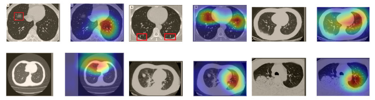
This paper explores how well deep learning models trained on chest CT images can diagnose COVID-19 infected people in a fast and automated process. To this end, we adopted advanced deep network architectures and proposed a transfer learning strategy using custom-sized input tailored for each deep architecture to achieve the best performance. We conducted extensive sets of experiments on two CT image datasets, namely, the SARS-CoV-2 CT-scan and the COVID19-CT. The results show superior performances for our models compared with previous studies. Our best models achieved average accuracy, precision, sensitivity, specificity, and F1-score values of 99.4%, 99.6%, 99.8%, 99.6%, and 99.4% on the SARS-CoV-2 dataset, and 92.9%, 91.3%, 93.7%, 92.2%, and 92.5% on the COVID19-CT dataset, respectively. For better interpretability of the results, we applied visualization techniques to provide visual explanations for the models' predictions. Feature visualizations of the learned features show well-separated clusters representing CT images of COVID-19 and non-COVID-19 cases. Moreover, the visualizations indicate that our models are not only capable of identifying COVID-19 cases but also provide accurate localization of the COVID-19-associated regions, as indicated by well-trained radiologists.

摘要

本文探讨了基于胸部 CT 图像训练的深度学习模型如何在快速自动化的过程中诊断 COVID-19 感染者。为此,我们采用了先进的深度网络架构,并提出了一种使用针对每个深度架构定制大小的输入的迁移学习策略,以实现最佳性能。我们在两个 CT 图像数据集上进行了广泛的实验,即 SARS-CoV-2 CT 扫描和 COVID19-CT。结果表明,与之前的研究相比,我们的模型表现出色。我们的最佳模型在 SARS-CoV-2 数据集上的平均准确率、精度、敏感度、特异性和 F1 得分为 99.4%、99.6%、99.8%、99.6%和 99.4%,在 COVID19-CT 数据集上的平均准确率、精度、敏感度、特异性和 F1 得分为 92.9%、91.3%、93.7%、92.2%和 92.5%。为了更好地解释结果,我们应用了可视化技术来为模型的预测提供可视化解释。学习特征的特征可视化显示了代表 COVID-19 和非 COVID-19 病例的 CT 图像的良好分离簇。此外,可视化结果表明,我们的模型不仅能够识别 COVID-19 病例,还能够提供 COVID-19 相关区域的准确定位,这与训练有素的放射科医生的指示一致。

https://cdn.ncbi.nlm.nih.gov/pmc/blobs/2ea9/7828058/10631070de05/sensors-21-00455-g018.jpg
https://cdn.ncbi.nlm.nih.gov/pmc/blobs/2ea9/7828058/3531d819c112/sensors-21-00455-g001.jpg
https://cdn.ncbi.nlm.nih.gov/pmc/blobs/2ea9/7828058/39333f3c6bd4/sensors-21-00455-g002.jpg
https://cdn.ncbi.nlm.nih.gov/pmc/blobs/2ea9/7828058/78587a8979c8/sensors-21-00455-g003.jpg
https://cdn.ncbi.nlm.nih.gov/pmc/blobs/2ea9/7828058/b9f560b7ad9c/sensors-21-00455-g004.jpg
https://cdn.ncbi.nlm.nih.gov/pmc/blobs/2ea9/7828058/537182b8ed75/sensors-21-00455-g005.jpg
https://cdn.ncbi.nlm.nih.gov/pmc/blobs/2ea9/7828058/0c3bee6900bc/sensors-21-00455-g006.jpg
https://cdn.ncbi.nlm.nih.gov/pmc/blobs/2ea9/7828058/90a9118fe563/sensors-21-00455-g007.jpg
https://cdn.ncbi.nlm.nih.gov/pmc/blobs/2ea9/7828058/0cc6129feaa6/sensors-21-00455-g008.jpg
https://cdn.ncbi.nlm.nih.gov/pmc/blobs/2ea9/7828058/48797f859095/sensors-21-00455-g009.jpg
https://cdn.ncbi.nlm.nih.gov/pmc/blobs/2ea9/7828058/a947d8e41f39/sensors-21-00455-g010.jpg
https://cdn.ncbi.nlm.nih.gov/pmc/blobs/2ea9/7828058/1c23c3adabf2/sensors-21-00455-g011.jpg
https://cdn.ncbi.nlm.nih.gov/pmc/blobs/2ea9/7828058/b68c98c5ad04/sensors-21-00455-g012.jpg
https://cdn.ncbi.nlm.nih.gov/pmc/blobs/2ea9/7828058/120669715037/sensors-21-00455-g013.jpg
https://cdn.ncbi.nlm.nih.gov/pmc/blobs/2ea9/7828058/d9f3f5824651/sensors-21-00455-g014.jpg
https://cdn.ncbi.nlm.nih.gov/pmc/blobs/2ea9/7828058/59e165dc2ebf/sensors-21-00455-g015.jpg
https://cdn.ncbi.nlm.nih.gov/pmc/blobs/2ea9/7828058/69a875a1290b/sensors-21-00455-g016.jpg
https://cdn.ncbi.nlm.nih.gov/pmc/blobs/2ea9/7828058/3231769981e5/sensors-21-00455-g017.jpg
https://cdn.ncbi.nlm.nih.gov/pmc/blobs/2ea9/7828058/10631070de05/sensors-21-00455-g018.jpg

相似文献

1
Explainable COVID-19 Detection Using Chest CT Scans and Deep Learning.利用胸部 CT 扫描和深度学习进行 COVID-19 的可解释检测。
Sensors (Basel). 2021 Jan 11;21(2):455. doi: 10.3390/s21020455.
2
Automated detection of COVID-19 using ensemble of transfer learning with deep convolutional neural network based on CT scans.基于 CT 扫描的深度学习卷积神经网络集成迁移学习的 COVID-19 自动检测。
Int J Comput Assist Radiol Surg. 2021 Jan;16(1):115-123. doi: 10.1007/s11548-020-02286-w. Epub 2020 Nov 16.
3
SOM-LWL method for identification of COVID-19 on chest X-rays.基于 SOM-LWL 算法的胸部 X 光片 COVID-19 识别方法。
PLoS One. 2021 Feb 24;16(2):e0247176. doi: 10.1371/journal.pone.0247176. eCollection 2021.
4
Detection and analysis of COVID-19 in medical images using deep learning techniques.使用深度学习技术在医学图像中检测和分析 COVID-19。
Sci Rep. 2021 Oct 4;11(1):19638. doi: 10.1038/s41598-021-99015-3.
5
Optimised genetic algorithm-extreme learning machine approach for automatic COVID-19 detection.优化遗传算法-极限学习机方法自动检测 COVID-19。
PLoS One. 2020 Dec 15;15(12):e0242899. doi: 10.1371/journal.pone.0242899. eCollection 2020.
6
Deep Learning-Based System Combining Chest X-Ray and Computerized Tomography Images for COVID-19 Diagnosis.基于深度学习的结合胸部 X 光和计算机断层扫描图像的 COVID-19 诊断系统。
Br J Hosp Med (Lond). 2024 Aug 30;85(8):1-15. doi: 10.12968/hmed.2024.0244. Epub 2024 Aug 27.
7
COVID-19 detection in CT and CXR images using deep learning models.使用深度学习模型进行 CT 和 CXR 图像中的 COVID-19 检测。
Biogerontology. 2022 Feb;23(1):65-84. doi: 10.1007/s10522-021-09946-7. Epub 2022 Jan 22.
8
Chest X-ray image phase features for improved diagnosis of COVID-19 using convolutional neural network.基于卷积神经网络的胸部 X 射线图像相位特征提高 COVID-19 诊断性能
Int J Comput Assist Radiol Surg. 2021 Feb;16(2):197-206. doi: 10.1007/s11548-020-02305-w. Epub 2021 Jan 9.
9
Classification of the COVID-19 infected patients using DenseNet201 based deep transfer learning.基于 DenseNet201 的深度迁移学习对 COVID-19 感染患者进行分类。
J Biomol Struct Dyn. 2021 Sep;39(15):5682-5689. doi: 10.1080/07391102.2020.1788642. Epub 2020 Jul 3.
10
An automated diagnosis and classification of COVID-19 from chest CT images using a transfer learning-based convolutional neural network.利用基于迁移学习的卷积神经网络对 chest CT 图像进行 COVID-19 的自动诊断和分类。
Comput Biol Med. 2022 May;144:105383. doi: 10.1016/j.compbiomed.2022.105383. Epub 2022 Mar 10.

引用本文的文献

1
Explainable AI for Oral Cancer Diagnosis: Multiclass Classification of Histopathology Images and Grad-CAM Visualization.用于口腔癌诊断的可解释人工智能:组织病理学图像的多类分类与Grad-CAM可视化
Biology (Basel). 2025 Jul 22;14(8):909. doi: 10.3390/biology14080909.
2
LGD_Net: Capsule network with extreme learning machine for classification of lung diseases using CT scans.LGD_Net:用于使用CT扫描对肺部疾病进行分类的带极限学习机的胶囊网络。
PLoS One. 2025 Aug 8;20(8):e0327419. doi: 10.1371/journal.pone.0327419. eCollection 2025.
3
A comparative analysis of sagittal, coronal, and axial magnetic resonance imaging planes in diagnosing anterior cruciate ligament and meniscal tears via a deep learning model: emphasizing the unexpected importance of the axial plane.

本文引用的文献

1
Deep neural network to detect COVID-19: one architecture for both CT Scans and Chest X-rays.用于检测新冠肺炎的深度神经网络:一种适用于CT扫描和胸部X光片的架构。
Appl Intell (Dordr). 2021;51(5):2777-2789. doi: 10.1007/s10489-020-01943-6. Epub 2020 Nov 6.
2
Deep Learning Enables Accurate Diagnosis of Novel Coronavirus (COVID-19) With CT Images.深度学习利用 CT 图像准确诊断新型冠状病毒(COVID-19)。
IEEE/ACM Trans Comput Biol Bioinform. 2021 Nov-Dec;18(6):2775-2780. doi: 10.1109/TCBB.2021.3065361. Epub 2021 Dec 8.
3
An Uncertainty-Aware Transfer Learning-Based Framework for COVID-19 Diagnosis.
通过深度学习模型对矢状面、冠状面和轴位磁共振成像平面在诊断前交叉韧带和半月板撕裂中的比较分析:强调轴位平面的意外重要性
Quant Imaging Med Surg. 2025 Jun 6;15(6):5811-5824. doi: 10.21037/qims-24-1808. Epub 2025 Jun 3.
4
Deep viewing for the identification of Covid-19 infection status from chest X-Ray image using CNN based architecture.基于卷积神经网络架构,通过深度观察从胸部X光图像识别新冠病毒感染状态。
Intell Syst Appl. 2022 Nov;16:200130. doi: 10.1016/j.iswa.2022.200130. Epub 2022 Oct 6.
5
Recent advances of nanotechnology in COVID 19: A critical review and future perspective.纳米技术在新型冠状病毒肺炎中的最新进展:批判性综述与未来展望
OpenNano. 2023 Jan;9:100118. doi: 10.1016/j.onano.2022.100118. Epub 2022 Dec 14.
6
A BiLSTM model enhanced with multi-objective arithmetic optimization for COVID-19 diagnosis from CT images.一种通过多目标算术优化增强的双向长短期记忆(BiLSTM)模型,用于从CT图像诊断新冠肺炎。
Sci Rep. 2025 Mar 29;15(1):10841. doi: 10.1038/s41598-025-94654-2.
7
Lung Ultrasonography in the Evaluation of Late Sequelae of COVID-19 Pneumonia-A Comparison with Chest Computed Tomography: A Prospective Study.肺部超声在评估 COVID-19 肺炎后遗症中的应用——与胸部 CT 的比较:一项前瞻性研究。
Viruses. 2024 Jun 3;16(6):905. doi: 10.3390/v16060905.
8
A comparative analysis of different augmentations for brain images.不同脑图像增强方法的比较分析。
Med Biol Eng Comput. 2024 Oct;62(10):3123-3150. doi: 10.1007/s11517-024-03127-7. Epub 2024 May 24.
9
DeepChestGNN: A Comprehensive Framework for Enhanced Lung Disease Identification through Advanced Graphical Deep Features.深胸图神经网络:通过先进的图形深度学习特征增强肺病识别的综合框架。
Sensors (Basel). 2024 Apr 29;24(9):2830. doi: 10.3390/s24092830.
10
AMTLDC: a new adversarial multi-source transfer learning framework to diagnosis of COVID-19.AMTLDC:一种用于新冠肺炎诊断的新型对抗多源迁移学习框架。
Evol Syst (Berl). 2023 Jan 12:1-15. doi: 10.1007/s12530-023-09484-2.
基于不确定性感知的迁移学习框架用于 COVID-19 诊断。
IEEE Trans Neural Netw Learn Syst. 2021 Apr;32(4):1408-1417. doi: 10.1109/TNNLS.2021.3054306. Epub 2021 Apr 2.
4
Deep learning for differentiating novel coronavirus pneumonia and influenza pneumonia.用于区分新型冠状病毒肺炎和流感肺炎的深度学习
Ann Transl Med. 2021 Jan;9(2):111. doi: 10.21037/atm-20-5328.
5
Rapid COVID-19 diagnosis using ensemble deep transfer learning models from chest radiographic images.使用来自胸部X光图像的集成深度迁移学习模型进行新冠病毒快速诊断。
J Ambient Intell Humaniz Comput. 2023;14(5):5541-5553. doi: 10.1007/s12652-020-02669-6. Epub 2020 Nov 16.
6
COVID-Net: a tailored deep convolutional neural network design for detection of COVID-19 cases from chest X-ray images.COVID-Net:一种针对胸部 X 光图像中 COVID-19 病例检测的定制化深度卷积神经网络设计。
Sci Rep. 2020 Nov 11;10(1):19549. doi: 10.1038/s41598-020-76550-z.
7
Deep learning-based model for detecting 2019 novel coronavirus pneumonia on high-resolution computed tomography.基于深度学习的高分辨率计算机断层扫描 2019 年新型冠状病毒肺炎检测模型。
Sci Rep. 2020 Nov 5;10(1):19196. doi: 10.1038/s41598-020-76282-0.
8
COVID-19 detection in CT images with deep learning: A voting-based scheme and cross-datasets analysis.基于深度学习的CT图像中COVID-19检测:一种基于投票的方案及跨数据集分析
Inform Med Unlocked. 2020;20:100427. doi: 10.1016/j.imu.2020.100427. Epub 2020 Sep 14.
9
Contrastive Cross-Site Learning With Redesigned Net for COVID-19 CT Classification.基于重新设计的网络的 COVID-19 CT 分类对比跨站点学习。
IEEE J Biomed Health Inform. 2020 Oct;24(10):2806-2813. doi: 10.1109/JBHI.2020.3023246. Epub 2020 Sep 10.
10
A Deep Learning System to Screen Novel Coronavirus Disease 2019 Pneumonia.一种用于筛查2019冠状病毒病肺炎的深度学习系统。
Engineering (Beijing). 2020 Oct;6(10):1122-1129. doi: 10.1016/j.eng.2020.04.010. Epub 2020 Jun 27.