National-Regional Key Technology Engineering Laboratory for Medical Ultrasound, Guangdong Key Laboratory for Biomedical Measurements and Ultrasound Imaging, Department of Biomedical Engineering, School of Medicine, Shenzhen University, Xueyuan Avenue, Nanshan District, Shenzhen, Guangdong Province, China.
Medical Imaging Department of Shenzhen Eye Hospital Affiliated to Jinan University, Shenzhen, Guangdong Province, China.
Transl Vis Sci Technol. 2020 Dec 9;9(2):61. doi: 10.1167/tvst.9.2.61. eCollection 2020 Dec.
To automate the segmentation of retinal layers, we propose DeepRetina, a method based on deep neural networks.
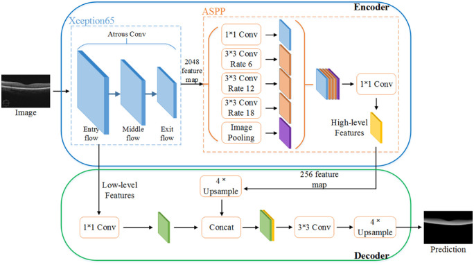
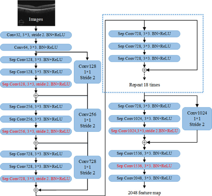
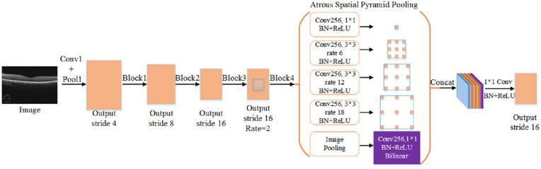
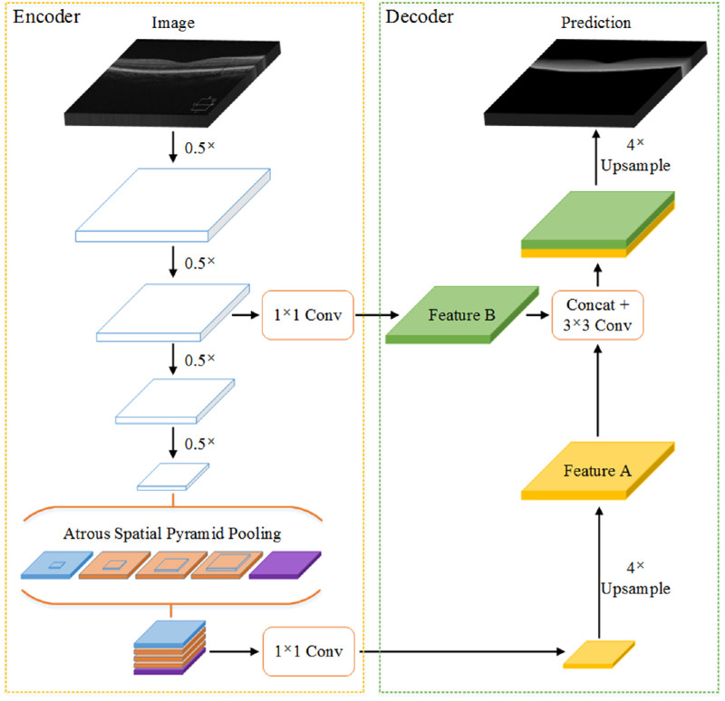
DeepRetina uses the improved Xception65 to extract and learn the characteristics of retinal layers. The Xception65-extracted feature maps are inputted to an atrous spatial pyramid pooling module to obtain multiscale feature information. This information is then recovered to capture clearer retinal layer boundaries in the encoder-decoder module, thus completing retinal layer auto-segmentation of the retinal optical coherence tomography (OCT) images.
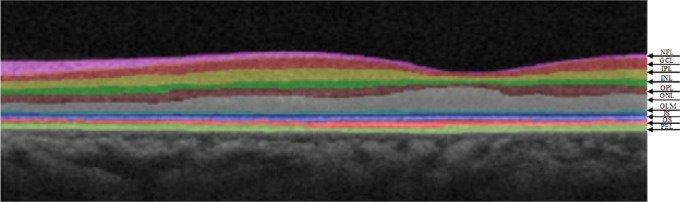
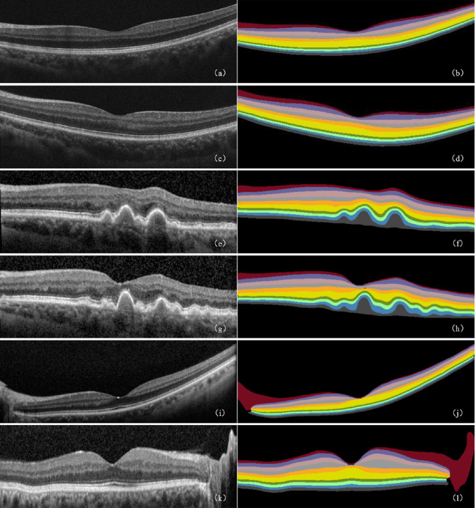
We validated this method using a retinal OCT image database containing 280 volumes (40 B-scans per volume) to demonstrate its effectiveness. The results showed that the method exhibits excellent performance in terms of the mean intersection over union and sensitivity (Se), which are as high as 90.41 and 92.15%, respectively. The intersection over union and Se values of the nerve fiber layer, ganglion cell layer, inner plexiform layer, inner nuclear layer, outer plexiform layer, outer nuclear layer, outer limiting membrane, photoreceptor inner segment, photoreceptor outer segment, and pigment epithelium layer were found to be above 88%.
DeepRetina can automate the segmentation of retinal layers and has great potential for the early diagnosis of fundus retinal diseases. In addition, our approach will provide a segmentation model framework for other types of tissues and cells in clinical practice.
Automating the segmentation of retinal layers can help effectively diagnose and monitor clinical retinal diseases. In addition, it requires only a small amount of manual segmentation, significantly improving work efficiency.
为实现视网膜层自动分割,我们提出了一种基于深度神经网络的方法 DeepRetina。
DeepRetina 使用改进的 Xception65 提取并学习视网膜层的特征。将 Xception65 提取的特征图输入空洞空间金字塔池化模块,以获取多尺度特征信息。然后,将该信息恢复到编码器-解码器模块中,以捕获更清晰的视网膜层边界,从而完成视网膜光学相干断层扫描(OCT)图像的视网膜层自动分割。
我们使用包含 280 个卷(每个卷 40 个 B 扫描)的视网膜 OCT 图像数据库验证了该方法的有效性。结果表明,该方法在平均交并比和敏感度(Se)方面表现出优异的性能,分别高达 90.41%和 92.15%。神经纤维层、节细胞层、内丛状层、内核层、外丛状层、外核层、外界膜、光感受器内节、光感受器外节和色素上皮层的交并比和 Se 值均高于 88%。
DeepRetina 可以自动分割视网膜层,对眼底视网膜疾病的早期诊断具有很大的潜力。此外,我们的方法将为临床实践中其他类型的组织和细胞提供分割模型框架。
杨可欣