Ohio State University Comprehensive Cancer Center, USA; Discovery Life Sciences, Powell, OH, USA.
Department of Pathology and Laboratory Medicine, Weill Cornell Medicine, NY, NY, USA.
Ann Diagn Pathol. 2021 Apr;51:151682. doi: 10.1016/j.anndiagpath.2020.151682. Epub 2020 Dec 24.
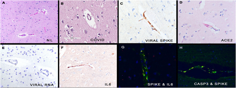
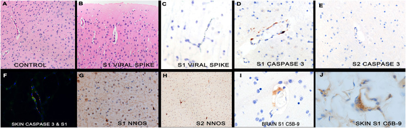
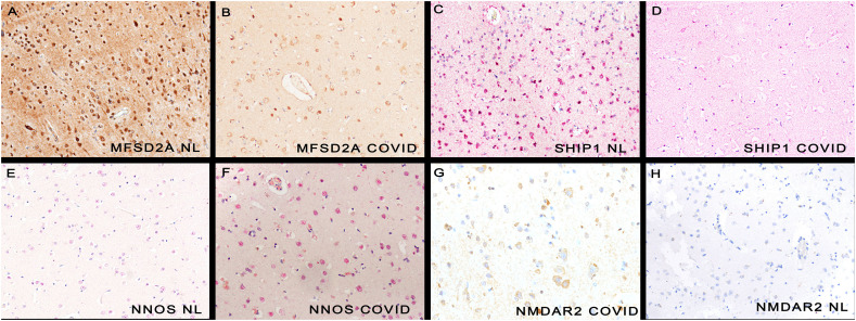
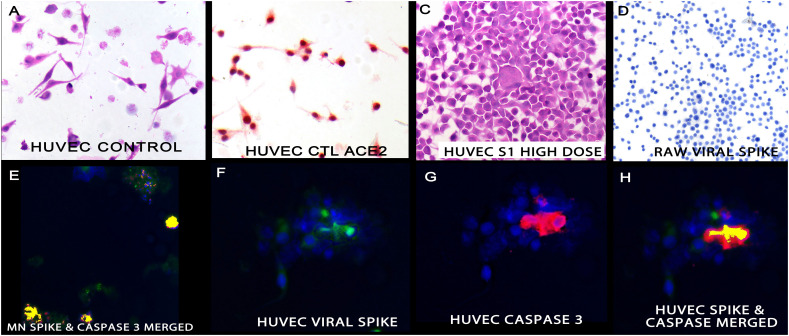
Neurologic complications of symptomatic COVID-19 are common. Brain tissues from 13 autopsies of people who died of COVID-19 were examined. Cultured endothelial and neuronal cells were incubated with and wild type mice were injected IV with different spike subunits. In situ analyses were used to detect SARS-CoV-2 proteins and the host response. In 13/13 brains from fatal COVID-19, pseudovirions (spike, envelope, and membrane proteins without viral RNA) were present in the endothelia of microvessels ranging from 0 to 14 positive cells/200× field (mean 4.3). The pseudovirions strongly co-localized with caspase-3, ACE2, IL6, TNFα, and C5b-9. The surrounding neurons demonstrated increased NMDAR2 and neuronal NOS plus decreased MFSD2a and SHIP1 proteins. Tail vein injection of the full length S1 spike subunit in mice led to neurologic signs (increased thirst, stressed behavior) not evident in those injected with the S2 subunit. The S1 subunit localized to the endothelia of microvessels in the mice brain and showed co-localization with caspase-3, ACE2, IL6, TNFα, and C5b-9. The surrounding neurons showed increased neuronal NOS and decreased MFSD2a. It is concluded that ACE2+ endothelial damage is a central part of SARS-CoV2 pathology and may be induced by the spike protein alone. Thus, the diagnostic pathologist can use either hematoxylin and eosin stain or immunohistochemistry for caspase 3 and ACE2 to document the endothelial cell damage of COVID-19.
新冠病毒感染的神经系统并发症较为常见。我们对 13 例死于新冠病毒感染的患者的脑组织进行了检查。我们培养了内皮细胞和神经元细胞,并用新冠病毒刺突蛋白的不同亚单位孵育这些细胞,并用这些亚单位对野生型小鼠进行静脉注射。我们利用原位分析技术来检测新冠病毒 S 蛋白和宿主反应。在 13 例死于新冠病毒感染的患者的脑组织中,我们发现了假病毒(无病毒 RNA 的刺突、包膜和膜蛋白),这些假病毒存在于微血管内皮细胞中,范围为 0 至 14 个阳性细胞/200×视野(平均值为 4.3)。这些假病毒与半胱天冬酶-3、血管紧张素转换酶 2(ACE2)、白细胞介素 6(IL6)、肿瘤坏死因子-α(TNFα)和 C5b-9 强烈共定位。周围神经元显示出 NMDAR2 增加和 MFSD2a、SHIP1 蛋白减少。在小鼠尾静脉注射全长 S1 刺突亚单位会导致神经症状(增加口渴、应激行为),而注射 S2 亚单位的小鼠则没有这些症状。S1 亚单位定位于小鼠大脑微血管内皮细胞,与半胱天冬酶-3、ACE2、IL6、TNFα 和 C5b-9 共定位。周围神经元显示出神经元 NOS 增加和 MFSD2a 减少。综上所述,ACE2+内皮损伤是新冠病毒 S 病理的核心部分,可能仅由刺突蛋白诱导。因此,病理诊断医生可以使用苏木精-伊红染色或 caspase 3 和 ACE2 的免疫组织化学染色来记录新冠病毒感染的内皮细胞损伤。