Skomorokhova Ekaterina A, Sankova Tatiana P, Orlov Iurii A, Savelev Andrew N, Magazenkova Daria N, Pliss Mikhail G, Skvortsov Alexey N, Sosnin Ilya M, Kirilenko Demid A, Grishchuk Ivan V, Sakhenberg Elena I, Polishchuk Elena V, Brunkov Pavel N, Romanov Alexey E, Puchkova Ludmila V, Ilyechova Ekaterina Yu
International Research Center of Functional Materials and Devices of Optoelectronics, ITMO University, St. Petersburg, Russia.
Department of Molecular Genetics, Research Institute of Experimental Medicine, St. Petersburg, Russia.
Nanotechnol Sci Appl. 2020 Dec 31;13:137-157. doi: 10.2147/NSA.S287658. eCollection 2020.
The ability of silver nanoparticles (AgNPs) of different sizes to influence copper metabolism in mice is assessed.
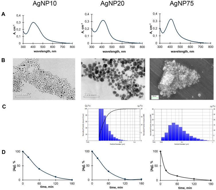
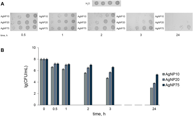
AgNPs with diameters of 10, 20, and 75 nm were fabricated through a chemical reduction of silver nitrate and characterized by UV/Vis spectrometry, transmission and scanning electronic microscopy, and laser diffractometry. To test their bioactivity, cells, cultured A549 cells, and C57Bl/6 mice were used. The antibacterial activity of AgNPs was determined by inhibition of colony-forming ability, and cytotoxicity was tested using the MTT test (viability, %). Ceruloplasmin (Cp, the major mammalian extracellular copper-containing protein) concentration and enzymatic activity were measured using gel-assay analyses and WB, respectively. In vitro binding of AgNPs with serum proteins was monitored with UV/Vis spectroscopy. Metal concentrations were measured using atomic absorption spectrometry.
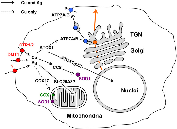
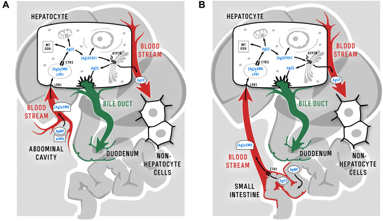
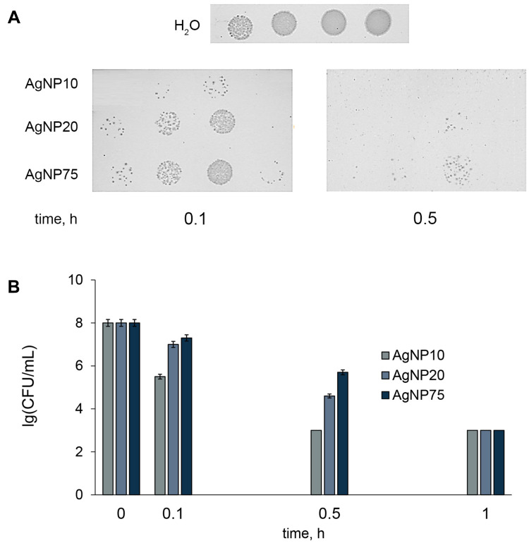
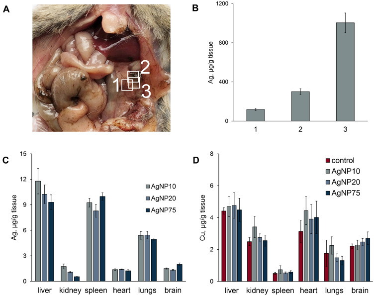
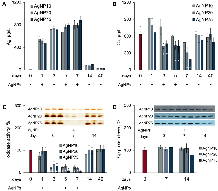
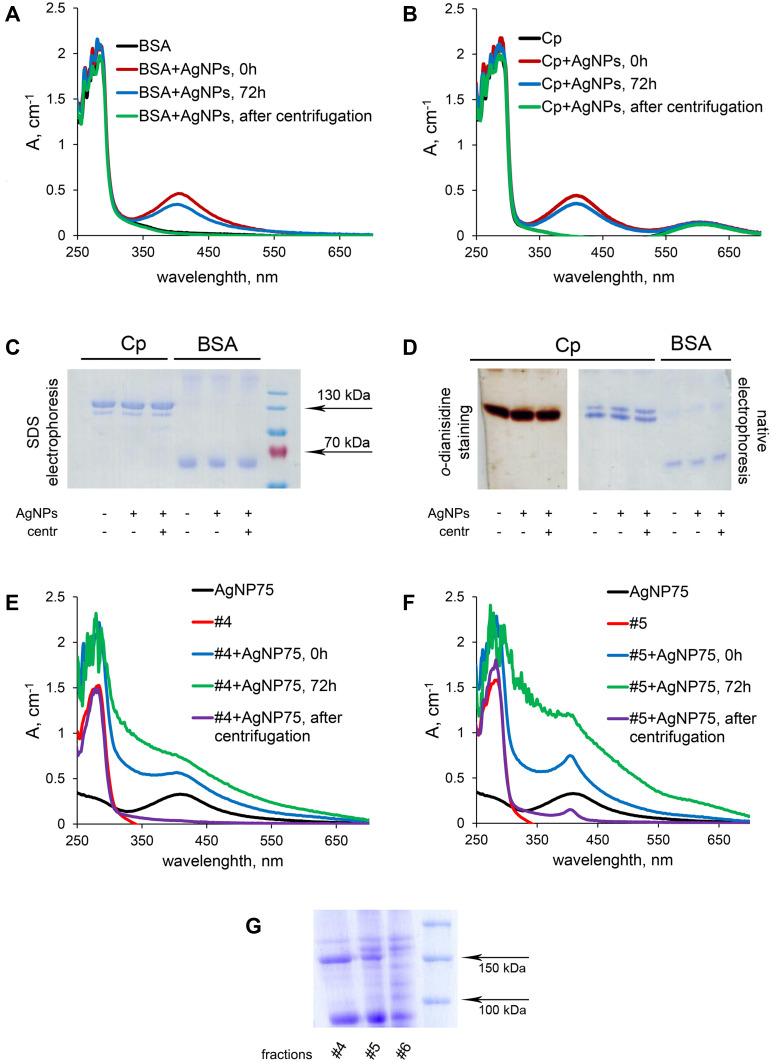
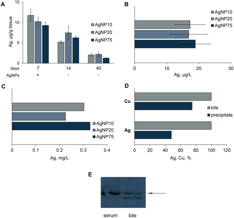
The smallest AgNPs displayed the largest dose- and time-dependent antibacterial activity. All nanoparticles inhibited the metabolic activity of A549 cells in accordance with dose and time, but no correlation between cytotoxicity and nanoparticle size was found. Nanosilver was not uniformly distributed through the body of mice intraperitoneally treated with low AgNP concentrations. It was predominantly accumulated in liver. There, nanosilver was included in ceruloplasmin, and Ag-ceruloplasmin with low oxidase activity level was formed. Larger nanoparticles more effectively interfered with the copper metabolism of mice. Large AgNPs quickly induced a drop of blood serum oxidase activity to practically zero, but after cancellation of AgNP treatment, the activity was rapidly restored. A major fraction of the nanosilver was excreted in the bile with Cp. Nanosilver was bound by alpha-2-macroglobulin in vitro and in vivo, but silver did not substitute for the copper atoms of Cp in vitro.
The data showed that even at low concentrations, AgNPs influence murine copper metabolism in size-dependent manner. This property negatively correlated with the antibacterial activity of AgNPs.
评估不同尺寸的银纳米颗粒(AgNPs)对小鼠铜代谢的影响能力。
通过硝酸银的化学还原制备直径为10、20和75nm的AgNPs,并通过紫外/可见光谱、透射和扫描电子显微镜以及激光衍射法进行表征。为了测试它们的生物活性,使用了细胞、培养的A549细胞和C57Bl/6小鼠。通过抑制菌落形成能力测定AgNPs的抗菌活性,并使用MTT试验(活力,%)测试细胞毒性。分别使用凝胶分析和WB测量铜蓝蛋白(Cp,主要的哺乳动物细胞外含铜蛋白)浓度和酶活性。用紫外/可见光谱监测AgNPs与血清蛋白的体外结合。使用原子吸收光谱法测量金属浓度。
最小的AgNPs表现出最大的剂量和时间依赖性抗菌活性。所有纳米颗粒均根据剂量和时间抑制A549细胞的代谢活性,但未发现细胞毒性与纳米颗粒尺寸之间的相关性。低浓度AgNP腹腔注射处理的小鼠体内,纳米银分布不均匀。它主要积聚在肝脏中。在那里,纳米银被纳入铜蓝蛋白,并形成了氧化酶活性水平较低的银 - 铜蓝蛋白。较大的纳米颗粒更有效地干扰小鼠的铜代谢。大的AgNPs迅速使血清氧化酶活性降至几乎为零,但在取消AgNP处理后,活性迅速恢复。大部分纳米银与Cp一起随胆汁排出。纳米银在体外和体内均与α-2-巨球蛋白结合,但在体外银不能替代铜蓝蛋白中的铜原子。
数据表明,即使在低浓度下,AgNPs也以尺寸依赖的方式影响小鼠的铜代谢。该特性与AgNPs的抗菌活性呈负相关。