Lee Dong Joon, Miguez Patricia, Kwon Jane, Daniel Renie, Padilla Ricardo, Min Samuel, Zalal Rahim, Ko Ching-Chang, Shin Hae Won
Oral and Craniofacial Health Science Institute, School of Dentistry, University of North Carolina, Chapel Hill, NC, USA.
Department of Periodontics, School of Dentistry, University of North Carolina, Chapel Hill, NC, USA.
J Tissue Eng. 2020 Dec 17;11:2041731420981672. doi: 10.1177/2041731420981672. eCollection 2020 Jan-Dec.
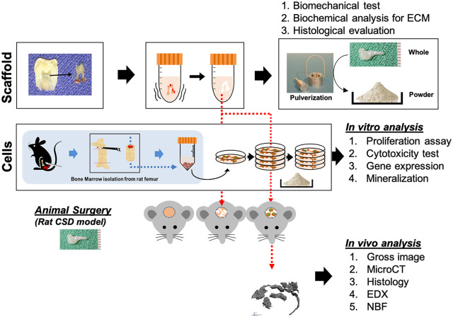
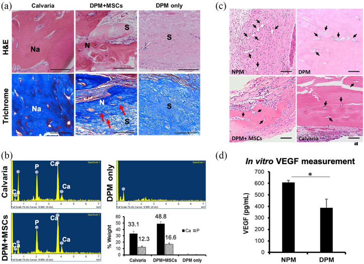
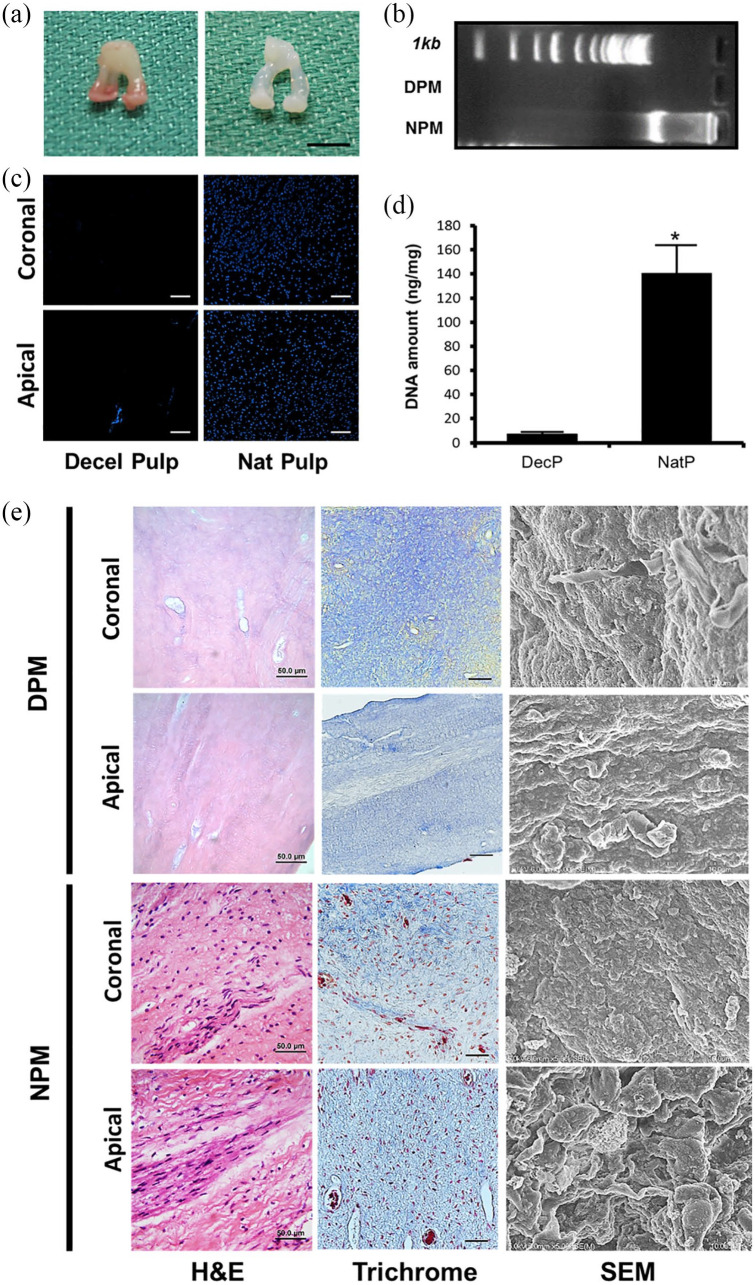
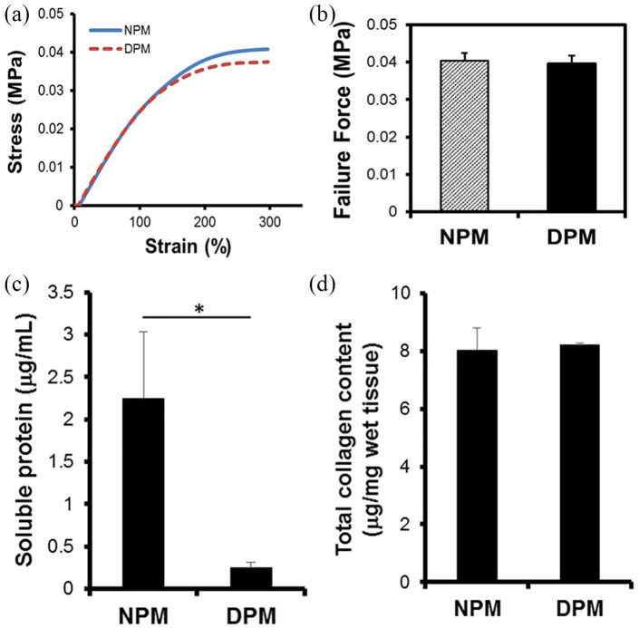
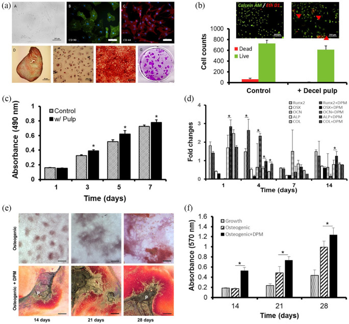
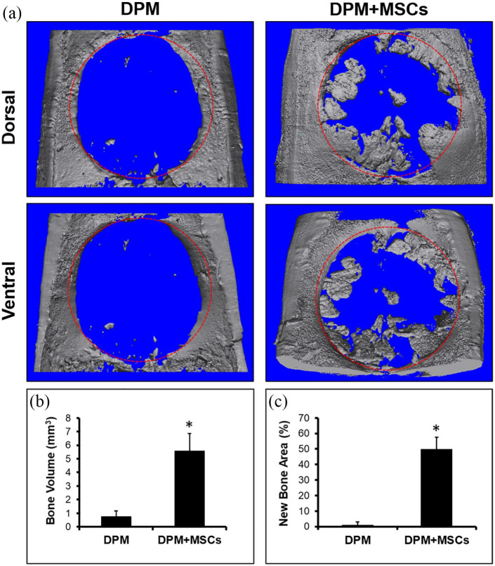
Scaffolds that are used for bone repair should provide an adequate environment for biomineralization by mesenchymal stem cells (MSCs). Recently, decellularized pulp matrices (DPM) have been utilized in endodontics for their high regenerative potential. Inspired by the dystrophic calcification on the pulp matrix known as pulp stone, we developed acellular pulp bioscaffolds and examined their potential in facilitating MSCs mineralization for bone defect repair. Pulp was decellularized, then retention of its structural integrity was confirmed by histological, mechanical, and biochemical evaluations. MSCs were seeded and proliferation, osteogenic gene expression, and biomineralization were assessed to verify DPM's osteogenic effects in vitro. MicroCT, energy-dispersive X-ray (EDX), and histological analyses were used to confirm that DPM seeded with MSCs result in greater mineralization on rat critical-sized defects than that without MSCs. Overall, our study proves DPM's potential to serve as a scaffolding material for MSC-mediated bone regeneration for future craniofacial bone tissue engineering.
用于骨修复的支架应为间充质干细胞(MSCs)的生物矿化提供适宜的环境。近来,脱细胞牙髓基质(DPM)因其高再生潜力而被应用于牙髓病学领域。受牙髓基质上称为牙髓石的营养不良性钙化启发,我们开发了脱细胞牙髓生物支架,并研究了其在促进MSCs矿化以修复骨缺损方面的潜力。牙髓经脱细胞处理,然后通过组织学、力学和生化评估来确认其结构完整性得以保留。接种MSCs后,评估其增殖、成骨基因表达和生物矿化情况,以验证DPM在体外的成骨作用。采用MicroCT、能量色散X射线(EDX)和组织学分析来证实,接种MSCs的DPM在大鼠临界尺寸骨缺损处比未接种MSCs的DPM能产生更多矿化。总体而言,我们的研究证明了DPM作为一种支架材料在未来颅面骨组织工程中用于MSCs介导的骨再生的潜力。