Institute for Synaptic Physiology, Center for Molecular Neurobiology Hamburg (ZMNH), University Medical Center Hamburg-Eppendorf, Falkenried 94, 20251, Hamburg, Germany.
J Neuroinflammation. 2021 Jan 10;18(1):21. doi: 10.1186/s12974-020-02048-0.
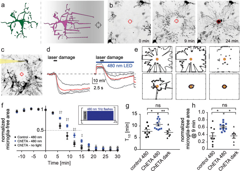
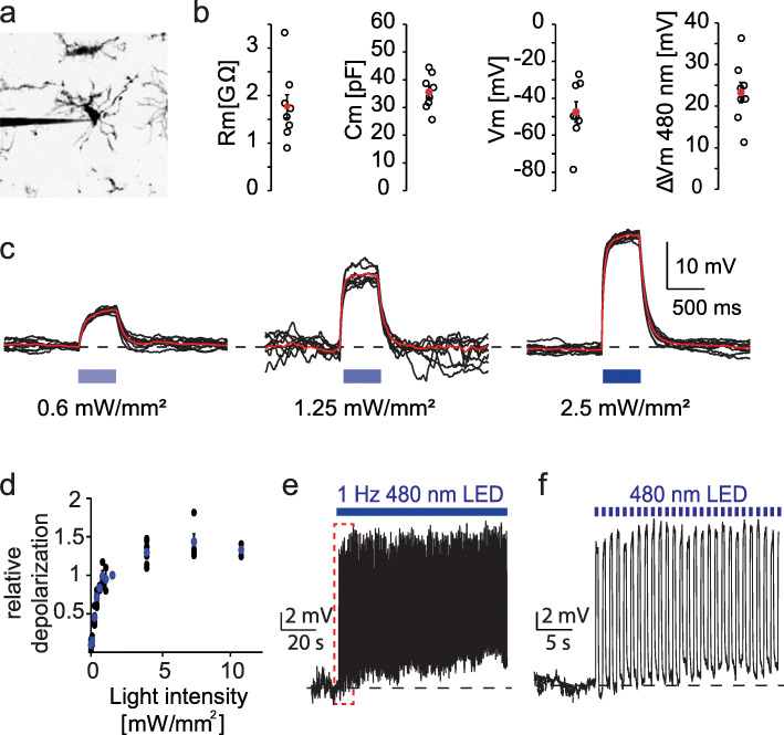
Microglia react to danger signals by rapid and targeted extension of cellular processes towards the source of the signal. This positive chemotactic response is accompanied by a hyperpolarization of the microglia membrane. Here, we show that optogenetic depolarization of microglia has little effect on baseline motility, but significantly slows down the chemotactic response. Reducing the extracellular Ca concentration mimics the effect of optogenetic depolarization. As the membrane potential sets the driving force for Ca entry, hyperpolarization is an integral part of rapid stimulus-response coupling in microglia. Compared to typical excitable cells such as neurons, the sign of the activating response is inverted in microglia, leading to inhibition by depolarizing channelrhodopsins.
小胶质细胞通过快速且靶向地将细胞过程延伸到信号源来对危险信号做出反应。这种正向趋化反应伴随着小胶质细胞膜的超极化。在这里,我们发现光遗传学去极化对小胶质细胞的基础运动几乎没有影响,但显著减缓了趋化反应。降低细胞外 Ca 浓度可以模拟光遗传学去极化的效果。由于细胞膜电位为 Ca 内流设定驱动力,因此超极化是小胶质细胞快速刺激-反应偶联的一个组成部分。与神经元等典型的可兴奋细胞相比,激活反应的符号在小胶质细胞中是反转的,导致去极化通道视紫红质的抑制作用。