• 文献检索
  • 文档翻译
  • 深度研究
  • 学术资讯
  • Suppr Zotero 插件Zotero 插件
  • 邀请有礼
  • 套餐&价格
  • 历史记录
应用&插件
Suppr Zotero 插件Zotero 插件浏览器插件Mac 客户端Windows 客户端微信小程序
定价
高级版会员购买积分包购买API积分包
服务
文献检索文档翻译深度研究API 文档MCP 服务
关于我们
关于 Suppr公司介绍联系我们用户协议隐私条款
关注我们

Suppr 超能文献

核心技术专利:CN118964589B侵权必究
粤ICP备2023148730 号-1Suppr @ 2026

文献检索

告别复杂PubMed语法,用中文像聊天一样搜索,搜遍4000万医学文献。AI智能推荐,让科研检索更轻松。

立即免费搜索

文件翻译

保留排版,准确专业,支持PDF/Word/PPT等文件格式,支持 12+语言互译。

免费翻译文档

深度研究

AI帮你快速写综述,25分钟生成高质量综述,智能提取关键信息,辅助科研写作。

立即免费体验

胸部计算机断层扫描(CT)在成人冠状病毒(Covid-19)肺炎检测和特征中的临床应用。

Clinical Application of Chest Computed Tomography (CT) in Detection and Characterization of Coronavirus (Covid-19) Pneumonia in Adults.

机构信息

Department of Radiology, Damietta Hospital, Al-Azhar University, Damietta, Egypt.

Czech Rehabilitation Hospital, Royal Health Group, Al-Ain, UAE.

出版信息

J Digit Imaging. 2021 Apr;34(2):273-283. doi: 10.1007/s10278-021-00426-5. Epub 2021 Feb 9.

DOI:10.1007/s10278-021-00426-5
PMID:33565000
原文链接:https://pmc.ncbi.nlm.nih.gov/articles/PMC7872307/
Abstract

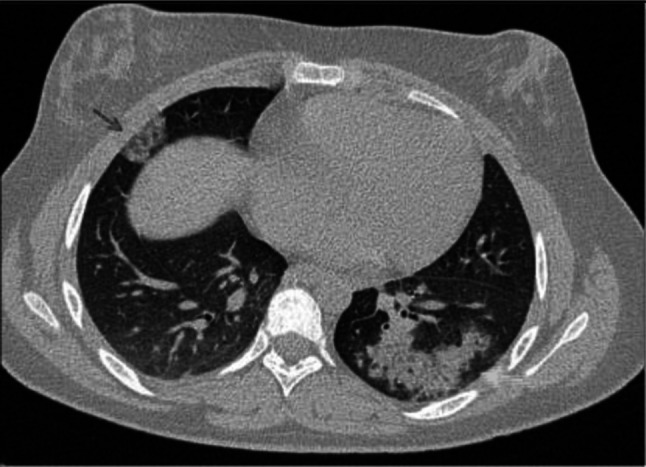
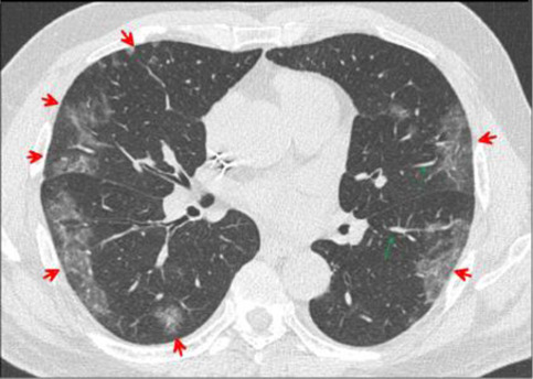
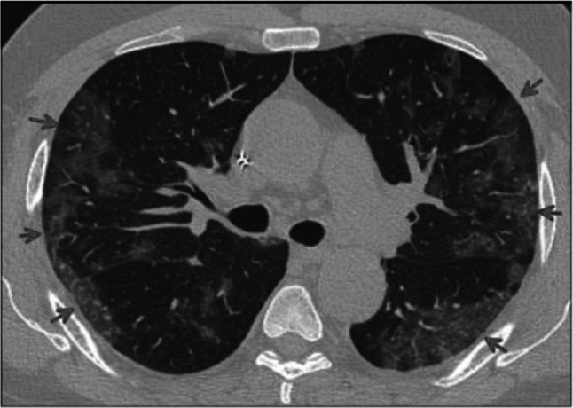
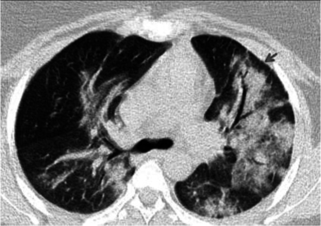
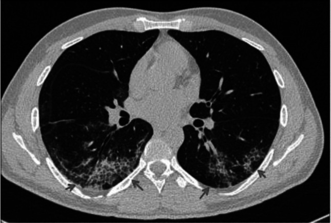
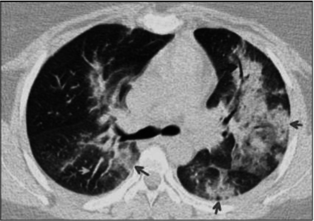
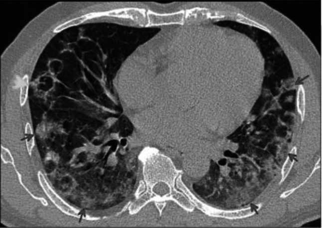
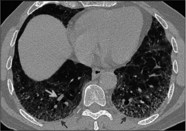
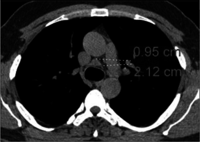
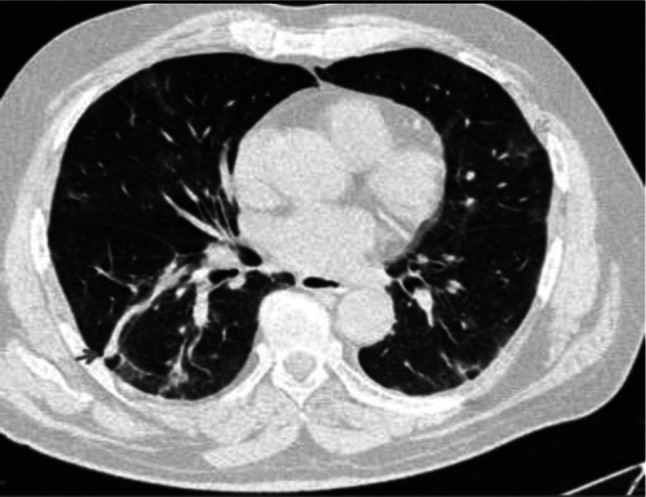
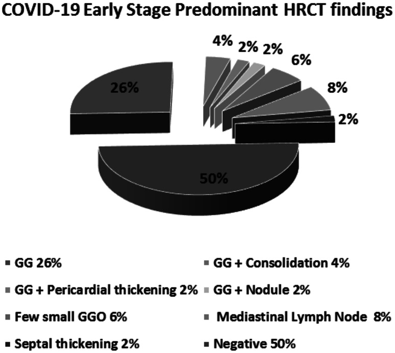
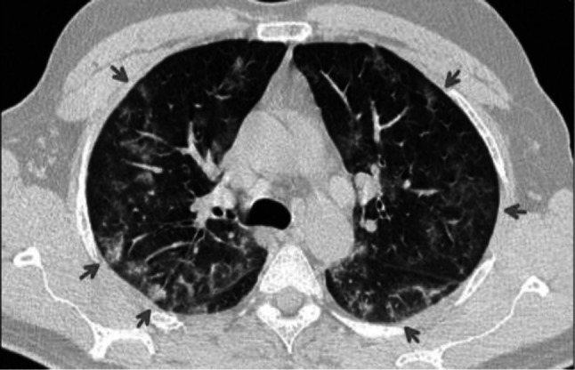
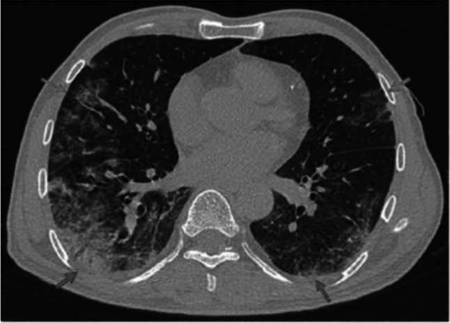
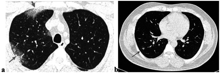
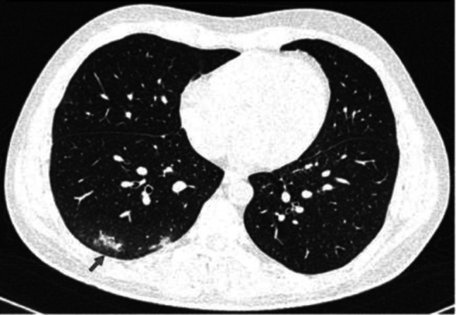
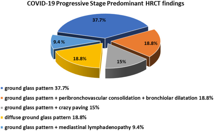
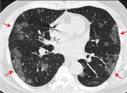
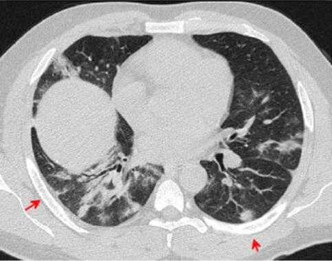
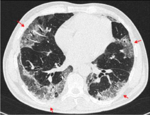
To analyze diagnostic accuracy of chest computed tomography (CT) and RT-PCR (real-time polymerase chain reaction) for COVID-19 (coronavirus disease 19) pneumonia in early and progressive stages. To evaluate if combination of chest CT with RT-PCR can supplement the shortage of RT-PCR in diagnosis of COVID-19 pneumonia. We conducted a prospective study on 103 male patients. The study population were divided into two groups; early COVID-19 stage (number = 50 patients, with positive RT-PCR but mild symptoms) and progressive COVID-19 stage (number = 53, positive RT-PCR and sever symptoms including fever > 37.5 °C, cough, and shortness of breath). All patients underwent CT imaging. The early stage included typical category; 34% (17 out of 50 cases), 6% indeterminate category (3 cases), 10% atypical category (5 cases) and 50% (25 cases) were normal CT imaging. The progressive stage included typical category that was further divided to five subgroups; (i) peripheral bilateral lower lobe ground-glass opacity (GGO) in (37.7%), (ii) peripheral bilateral lower lobes GGO with peribronchovascular consolidation and bronchiolar dilatation in (18.8%), (iii) peripheral bilateral lower lobes GGO with crazy paving appearance in (15%), (iv) bilateral diffuse GGO in (18.8%), and (v) peripheral bilateral GGO with mediastinal lymph node enlargement (9.4%). Chest CT imaging could aid to supplement the shortages of PCR for clinically suspected patients of COVID-19 in the epidemic area as CT was positive in 50% of patients. Chest CT is very effective in detecting pulmonary parenchymal abnormalities in the progressive stage of COVID-19 patients in 100%.

摘要

分析胸部计算机断层扫描(CT)和实时聚合酶链反应(RT-PCR)在 COVID-19(冠状病毒病 19)肺炎早期和进展期的诊断准确性。评估胸部 CT 与 RT-PCR 联合应用是否可以弥补 RT-PCR 在 COVID-19 肺炎诊断中的不足。我们对 103 名男性患者进行了前瞻性研究。研究人群分为两组;早期 COVID-19 期(阳性 RT-PCR 但症状较轻,病例数=50 例)和进展期 COVID-19 期(阳性 RT-PCR 且症状严重,包括发热>37.5°C、咳嗽和呼吸急促,病例数=53 例)。所有患者均行 CT 成像。早期阶段包括典型类别;34%(50 例中的 17 例)、6%不确定类别(3 例)、10%非典型类别(5 例)和 50%(25 例)为正常 CT 成像。进展期包括进一步分为五个亚组的典型类别;(i)外周双侧下叶磨玻璃密度影(GGO)(37.7%),(ii)外周双侧下叶 GGO 伴支气管血管周围实变和细支气管扩张(18.8%),(iii)外周双侧下叶 GGO 伴铺路石样外观(15%),(iv)双侧弥漫性 GGO(18.8%)和(v)外周双侧 GGO 伴纵隔淋巴结肿大(9.4%)。在疫情地区,胸部 CT 成像可辅助补充 PCR 对临床疑似 COVID-19 患者的不足,因为 50%的患者 CT 呈阳性。胸部 CT 在 COVID-19 患者进展期检测肺实质异常非常有效,准确率为 100%。

https://cdn.ncbi.nlm.nih.gov/pmc/blobs/f912/8290009/12212370ecce/10278_2021_426_Fig19_HTML.jpg
https://cdn.ncbi.nlm.nih.gov/pmc/blobs/f912/8290009/c6f595545985/10278_2021_426_Fig1_HTML.jpg
https://cdn.ncbi.nlm.nih.gov/pmc/blobs/f912/8290009/71fc6630c044/10278_2021_426_Fig2_HTML.jpg
https://cdn.ncbi.nlm.nih.gov/pmc/blobs/f912/8290009/4647a0fb16d1/10278_2021_426_Fig3_HTML.jpg
https://cdn.ncbi.nlm.nih.gov/pmc/blobs/f912/8290009/904e5fa5ee52/10278_2021_426_Fig4_HTML.jpg
https://cdn.ncbi.nlm.nih.gov/pmc/blobs/f912/8290009/9f02d84be253/10278_2021_426_Fig5_HTML.jpg
https://cdn.ncbi.nlm.nih.gov/pmc/blobs/f912/8290009/3cc95931642b/10278_2021_426_Fig6_HTML.jpg
https://cdn.ncbi.nlm.nih.gov/pmc/blobs/f912/8290009/716cefb06c28/10278_2021_426_Fig7_HTML.jpg
https://cdn.ncbi.nlm.nih.gov/pmc/blobs/f912/8290009/7ad4fee54ed5/10278_2021_426_Fig8_HTML.jpg
https://cdn.ncbi.nlm.nih.gov/pmc/blobs/f912/8290009/7758b2a75b48/10278_2021_426_Fig9_HTML.jpg
https://cdn.ncbi.nlm.nih.gov/pmc/blobs/f912/8290009/582f0043f0e1/10278_2021_426_Fig10_HTML.jpg
https://cdn.ncbi.nlm.nih.gov/pmc/blobs/f912/8290009/3ae118abab97/10278_2021_426_Fig11_HTML.jpg
https://cdn.ncbi.nlm.nih.gov/pmc/blobs/f912/8290009/18952da01a6f/10278_2021_426_Fig12_HTML.jpg
https://cdn.ncbi.nlm.nih.gov/pmc/blobs/f912/8290009/b97a5f338778/10278_2021_426_Fig13_HTML.jpg
https://cdn.ncbi.nlm.nih.gov/pmc/blobs/f912/8290009/77f98d69998d/10278_2021_426_Fig14_HTML.jpg
https://cdn.ncbi.nlm.nih.gov/pmc/blobs/f912/8290009/b23df1a8296f/10278_2021_426_Fig15_HTML.jpg
https://cdn.ncbi.nlm.nih.gov/pmc/blobs/f912/8290009/f658e89a37ad/10278_2021_426_Fig16_HTML.jpg
https://cdn.ncbi.nlm.nih.gov/pmc/blobs/f912/8290009/ffd4f273d726/10278_2021_426_Fig17_HTML.jpg
https://cdn.ncbi.nlm.nih.gov/pmc/blobs/f912/8290009/b60cccc25be5/10278_2021_426_Fig18_HTML.jpg
https://cdn.ncbi.nlm.nih.gov/pmc/blobs/f912/8290009/12212370ecce/10278_2021_426_Fig19_HTML.jpg

相似文献

1
Clinical Application of Chest Computed Tomography (CT) in Detection and Characterization of Coronavirus (Covid-19) Pneumonia in Adults.胸部计算机断层扫描(CT)在成人冠状病毒(Covid-19)肺炎检测和特征中的临床应用。
J Digit Imaging. 2021 Apr;34(2):273-283. doi: 10.1007/s10278-021-00426-5. Epub 2021 Feb 9.
2
Temporal relationship between serial RT-PCR results and serial chest CT imaging, and serial CT changes in coronavirus 2019 (COVID-19) pneumonia: a descriptive study of 155 cases in China.新型冠状病毒(COVID-19)肺炎的实时逆转录聚合酶链反应(RT-PCR)结果与系列胸部 CT 影像学及系列 CT 改变的时间关系:中国 155 例的描述性研究。
Eur Radiol. 2021 Mar;31(3):1175-1184. doi: 10.1007/s00330-020-07268-9. Epub 2020 Sep 15.
3
Spectrum of chest computed tomographic (CT) findings in coronavirus disease-19 (COVID-19) patients in India.印度 COVID-19 患者的胸部计算机断层扫描(CT)表现谱。
Eur J Radiol. 2020 Aug;129:109147. doi: 10.1016/j.ejrad.2020.109147. Epub 2020 Jun 24.
4
CT imaging features of 34 patients infected with COVID-19.34 例新冠病毒感染患者的 CT 影像学特征。
Clin Imaging. 2020 Dec;68:226-231. doi: 10.1016/j.clinimag.2020.05.016. Epub 2020 May 16.
5
Diagnostic performance of chest CT to differentiate COVID-19 pneumonia in non-high-epidemic area in Japan.日本非高流行地区胸部CT鉴别新型冠状病毒肺炎的诊断性能
Jpn J Radiol. 2020 May;38(5):400-406. doi: 10.1007/s11604-020-00958-w. Epub 2020 Mar 30.
6
Chest CT findings in RT-PCR positive asymptomatic COVID-19 patients.胸部 CT 影像学表现与 RT-PCR 阳性无症状 COVID-19 患者。
Clin Imaging. 2021 Sep;77:37-42. doi: 10.1016/j.clinimag.2021.01.030. Epub 2021 Jan 30.
7
Imaging and clinical features of patients with 2019 novel coronavirus SARS-CoV-2.新型冠状病毒 2019 年 SARS-CoV-2 患者的影像学和临床特征。
Eur J Nucl Med Mol Imaging. 2020 May;47(5):1275-1280. doi: 10.1007/s00259-020-04735-9. Epub 2020 Feb 28.
8
[Spatial and temporal distribution and predictive value of chest CT scoring in patients with COVID-19].新型冠状病毒肺炎患者胸部CT评分的时空分布及预测价值
Zhonghua Jie He He Hu Xi Za Zhi. 2021 Mar 12;44(3):230-236. doi: 10.3760/cma.j.cn112147-20200522-00626.
9
Clinical Features and Chest CT Manifestations of Coronavirus Disease 2019 (COVID-19) in a Single-Center Study in Shanghai, China.中国上海单中心研究的 2019 年冠状病毒病(COVID-19)的临床特征和胸部 CT 表现。
AJR Am J Roentgenol. 2020 Jul;215(1):121-126. doi: 10.2214/AJR.20.22959. Epub 2020 Mar 14.
10
Comparison of the computed tomography findings in COVID-19 and other viral pneumonia in immunocompetent adults: a systematic review and meta-analysis.COVID-19 与免疫功能正常成年人其他病毒性肺炎的计算机断层扫描表现比较:系统评价和荟萃分析。
Eur Radiol. 2020 Dec;30(12):6485-6496. doi: 10.1007/s00330-020-07018-x. Epub 2020 Jun 27.

引用本文的文献

1
Phantom-based evaluation of image quality in Transformer-enhanced 2048-matrix CT imaging at low and ultralow doses.基于体模的低剂量和超低剂量Transformer增强型2048矩阵CT成像图像质量评估
Jpn J Radiol. 2025 Apr 7. doi: 10.1007/s11604-025-01755-z.
2
A Survey on AI Techniques for Thoracic Diseases Diagnosis Using Medical Images.基于医学图像的胸部疾病诊断人工智能技术综述
Diagnostics (Basel). 2022 Dec 3;12(12):3034. doi: 10.3390/diagnostics12123034.
3
Update on the limited sensitivity of computed tomography relative to RT-PCR for COVID-19: a systematic review.

本文引用的文献

1
Radiological Society of North America Expert Consensus Document on Reporting Chest CT Findings Related to COVID-19: Endorsed by the Society of Thoracic Radiology, the American College of Radiology, and RSNA.北美放射学会关于报告与COVID-19相关的胸部CT检查结果的专家共识文件:得到了胸腔放射学会、美国放射学会和北美放射学会的认可。
Radiol Cardiothorac Imaging. 2020 Mar 25;2(2):e200152. doi: 10.1148/ryct.2020200152. eCollection 2020 Apr.
2
Coronavirus Disease 2019: Initial Detection on Chest CT in a Retrospective Multicenter Study of 103 Chinese Patients.2019冠状病毒病:103例中国患者回顾性多中心研究中胸部CT的初步检测
Radiol Cardiothorac Imaging. 2020 Apr 6;2(2):e200092. doi: 10.1148/ryct.2020200092. eCollection 2020 Apr.
3
计算机断层扫描相对于新冠病毒逆转录聚合酶链反应检测的敏感性有限的最新情况:一项系统评价
Pol J Radiol. 2022 Jul 12;87:e381-e391. doi: 10.5114/pjr.2022.118238. eCollection 2022.
4
Benign incidental cardiac findings in chest and cardiac CT imaging.胸部和心脏 CT 成像中的良性偶然心脏发现。
Br J Radiol. 2023 Feb;96(1142):20211302. doi: 10.1259/bjr.20211302. Epub 2022 Aug 22.
5
Detection of COVID-19 from voice, cough and breathing patterns: Dataset and preliminary results.从声音、咳嗽和呼吸模式中检测新冠病毒:数据集与初步结果
Comput Biol Med. 2021 Nov;138:104944. doi: 10.1016/j.compbiomed.2021.104944. Epub 2021 Oct 13.
6
The Imaging Informatics Response to a Pandemic.影像信息学对大流行病的应对。
J Digit Imaging. 2021 Apr;34(2):229-230. doi: 10.1007/s10278-021-00445-2.
Recommendations for coronavirus disease 2019 (COVID-19) prevention and infection control in the radiology department: Chinese experience.新型冠状病毒肺炎(COVID-19)放射科预防与感染控制建议:中国经验。
Clin Imaging. 2021 Jan;69:33-36. doi: 10.1016/j.clinimag.2020.06.044. Epub 2020 Jul 1.
4
Interpreting a covid-19 test result.解读新冠病毒检测结果。
BMJ. 2020 May 12;369:m1808. doi: 10.1136/bmj.m1808.
5
Diagnostic performance between CT and initial real-time RT-PCR for clinically suspected 2019 coronavirus disease (COVID-19) patients outside Wuhan, China.中国武汉以外地区临床疑似 2019 冠状病毒病(COVID-19)患者的 CT 与实时 RT-PCR 检测的诊断性能比较。
Respir Med. 2020 Jul;168:105980. doi: 10.1016/j.rmed.2020.105980. Epub 2020 Apr 21.
6
Mediastinal lymphadenopathy in patients with severe COVID-19.重症新型冠状病毒肺炎患者的纵隔淋巴结肿大
Lancet Infect Dis. 2020 Nov;20(11):1230. doi: 10.1016/S1473-3099(20)30310-8. Epub 2020 Apr 21.
7
Coronavirus Disease 2019 (COVID-19) CT Findings: A Systematic Review and Meta-analysis.新型冠状病毒病 2019(COVID-19)的 CT 表现:系统评价和荟萃分析。
J Am Coll Radiol. 2020 Jun;17(6):701-709. doi: 10.1016/j.jacr.2020.03.006. Epub 2020 Mar 25.
8
CT features of SARS-CoV-2 pneumonia according to clinical presentation: a retrospective analysis of 120 consecutive patients from Wuhan city.根据临床表现的 SARS-CoV-2 肺炎的 CT 特征:对武汉市 120 例连续患者的回顾性分析。
Eur Radiol. 2020 Aug;30(8):4417-4426. doi: 10.1007/s00330-020-06854-1. Epub 2020 Apr 11.
9
Pathogenesis of COVID-19 from a cell biology perspective.从细胞生物学角度看 COVID-19 的发病机制。
Eur Respir J. 2020 Apr 16;55(4). doi: 10.1183/13993003.00607-2020. Print 2020 Apr.
10
Understanding COVID-19: what does viral RNA load really mean?了解新冠病毒:病毒RNA载量究竟意味着什么?
Lancet Infect Dis. 2020 Jun;20(6):635-636. doi: 10.1016/S1473-3099(20)30237-1. Epub 2020 Mar 27.