Department of Neurobiology, University of Pittsburgh, Pittsburgh, Pennsylvania, USA.
Institute of Neuroscience, Center for Excellence in Brain Science and Intelligence Technology, Chinese Academy of Sciences, Shanghai, China.
ILAR J. 2020 Dec 31;61(2-3):274-285. doi: 10.1093/ilar/ilaa029.
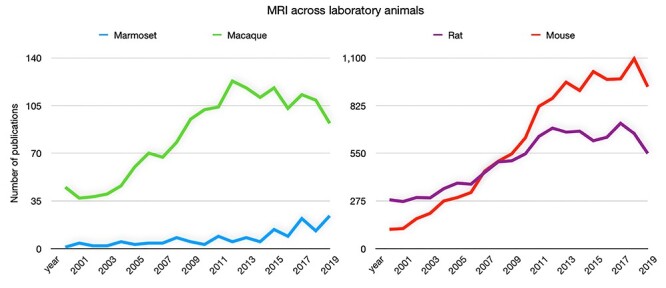
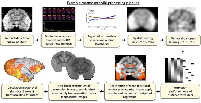
The use of the common marmoset monkey (Callithrix jacchus) for neuroscientific research has grown markedly in the last decade. Magnetic resonance imaging (MRI) has played a significant role in establishing the extent of comparability of marmoset brain architecture with the human brain and brains of other preclinical species (eg, macaques and rodents). As a non-invasive technique, MRI allows for the flexible acquisition of the same sequences across different species in vivo, including imaging of whole-brain functional topologies not possible with more invasive techniques. Being one of the smallest New World primates, the marmoset may be an ideal nonhuman primate species to study with MRI. As primates, marmosets have an elaborated frontal cortex with features analogous to the human brain, while also having a small enough body size to fit into powerful small-bore MRI systems typically employed for rodent imaging; these systems offer superior signal strength and resolution. Further, marmosets have a rich behavioral repertoire uniquely paired with a lissencephalic cortex (like rodents). This smooth cortical surface lends itself well to MRI and also other invasive methodologies. With the advent of transgenic modification techniques, marmosets have gained significant traction as a powerful complement to canonical mammalian modelling species. Marmosets are poised to make major contributions to preclinical investigations of the pathophysiology of human brain disorders as well as more basic mechanistic explorations of the brain. The goal of this article is to provide an overview of the practical aspects of implementing MRI and fMRI in marmosets (both under anesthesia and fully awake) and discuss the development of resources recently made available for marmoset imaging.
在过去的十年中,普通狨猴(Callithrix jacchus)在神经科学研究中的应用显著增加。磁共振成像(MRI)在确定狨猴大脑结构与人类大脑和其他临床前物种(如猕猴和啮齿动物)大脑的可比性方面发挥了重要作用。作为一种非侵入性技术,MRI 允许在体内灵活地获取不同物种的相同序列,包括使用更具侵入性的技术无法进行的全脑功能拓扑成像。作为新世界中最小的灵长类动物之一,狨猴可能是用 MRI 进行研究的理想非人类灵长类动物。作为灵长类动物,狨猴具有与人类大脑类似的复杂额叶皮层,同时其体型足够小,可以放入通常用于啮齿动物成像的大功率小口径 MRI 系统中;这些系统提供了更高的信号强度和分辨率。此外,狨猴具有丰富的行为 repertoire,与脑回较少的大脑(如啮齿动物)独特配对。这种光滑的皮质表面非常适合 MRI 以及其他侵入性方法。随着转基因修饰技术的出现,狨猴作为经典哺乳动物模型物种的有力补充得到了显著发展。狨猴有望为人类大脑疾病的病理生理学的临床前研究以及大脑的更基本机制探索做出重大贡献。本文的目的是概述在狨猴(麻醉和完全清醒状态下)中实施 MRI 和 fMRI 的实用方面,并讨论最近为狨猴成像提供的资源的发展。