Department of Radiology, IMSUT Hospital, The Institute of Medical Science, The University of Tokyo.
Department of Radiology, The University of Tokyo Hospital.
Magn Reson Med Sci. 2022 Mar 1;21(1):95-109. doi: 10.2463/mrms.rev.2020-0159. Epub 2021 Mar 10.
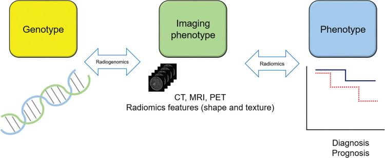
Texture analysis, as well as its broader category radiomics, describes a variety of techniques for image analysis that quantify the variation in surface intensity or patterns, including some that are imperceptible to the human visual system. Cerebral gliomas have been most rigorously studied in brain tumors using MR-based texture analysis (MRTA) to determine the correlation of various clinical measures with MRTA features. Promising results in cerebral gliomas have been shown in the previous MRTA studies in terms of the correlation with the World Health Organization grades, risk stratification in gliomas, and the differentiation of gliomas from other brain tumors. Multiple MRTA studies in gliomas have repeatedly shown high performance of entropy, a measure of the randomness in image intensity values, of either histogram- or gray-level co-occurrence matrix parameters. Similarly, researchers have applied MRTA to other brain tumors, including meningiomas and pediatric posterior fossa tumors.However, the value of MRTA in the clinical use remains undetermined, probably because previous studies have shown only limited reproducibility of the result in the real world. The low-to-modest generalizability may be attributed to variations in MRTA methods, sampling bias that originates from single-institution studies, and overfitting problems to a limited number of samples.To enhance the reliability and reproducibility of MRTA studies, researchers have realized the importance of standardizing methods in the field of radiomics. Another advancement is the recent development of a comprehensive assessment system to ensure the quality of a radiomics study. These two-way approaches will secure the validity of upcoming MRTA studies. The clinical use of texture analysis in brain MRI will be accelerated by these continuous efforts.
纹理分析及其更广泛的范畴放射组学描述了各种用于分析图像的技术,这些技术可以量化表面强度或模式的变化,包括一些人类视觉系统无法察觉的变化。脑胶质瘤是在脑肿瘤中使用基于磁共振的纹理分析(MRTA)进行研究最严格的肿瘤,以确定各种临床指标与 MRTA 特征的相关性。在以前的 MRTA 研究中,脑胶质瘤在与世界卫生组织分级、胶质瘤风险分层以及胶质瘤与其他脑肿瘤的鉴别方面显示出了有希望的结果。多项脑胶质瘤的 MRTA 研究反复表明,熵的性能很高,熵是图像强度值随机性的一种度量,无论是直方图还是灰度共生矩阵参数都可以衡量熵。同样,研究人员也将 MRTA 应用于其他脑肿瘤,包括脑膜瘤和儿童后颅窝肿瘤。然而,MRTA 在临床应用中的价值仍未确定,可能是因为之前的研究仅显示出在现实世界中结果的可重复性有限。这种低到中等的泛化能力可能归因于 MRTA 方法的变化、源于单机构研究的采样偏差以及对有限数量的样本过度拟合的问题。为了提高 MRTA 研究的可靠性和可重复性,研究人员已经意识到在放射组学领域标准化方法的重要性。另一个进展是最近开发了一个全面的评估系统,以确保放射组学研究的质量。这两种方法都将确保即将进行的 MRTA 研究的有效性。通过这些持续的努力,纹理分析在脑 MRI 中的临床应用将得到加速。