Mireles Marcela, Soule Cody W, Dehghani Mehdi, Gaborski Thomas R
Department of Biomedical Engineering, Rochester Institute of Technology, Rochester, NY, USA.
Department of Biomedical Engineering, University of Rochester, Rochester, NY, USA.
Nanoscale Adv. 2020 Oct;2(10):4427-4436. doi: 10.1039/D0NA00142B. Epub 2020 May 12.
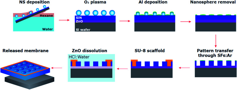
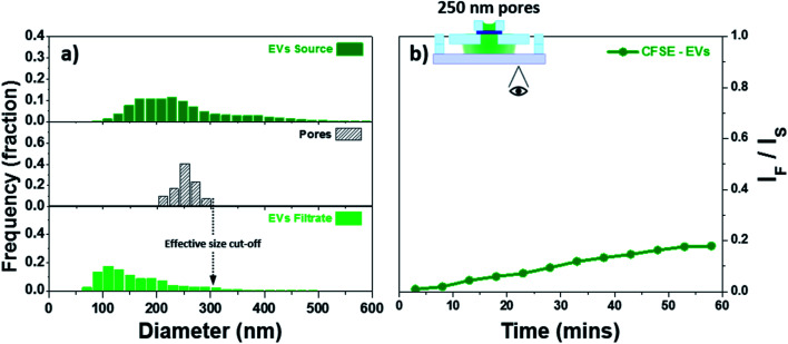
Nanoscale biocomponents naturally released by cells, such as extracellular vesicles (EVs), have recently gained interest due to their therapeutic and diagnostic potential. Membrane based isolation and co-culture systems have been utilized in an effort to study EVs and their effects. Nevertheless, improved platforms for the study of small EVs are still needed. Suitable membranes, for isolation and co-culture systems, require pore sizes to reach into the nanoscale. These pore sizes cannot be achieved through traditional lithographic techniques and conventional thick nanoporous membranes commonly exhibit low permeability. Here we utilized nanospheres, similar in size and shape to the targeted small EVs, as patterning features for the fabrication of freestanding SiN membranes (120 nm thick) released in minutes through a sacrificial ZnO layer. We evaluated the feasibility of separating subpopulation of EVs based on size using these membranes. The membrane used here showed an effective size cut-off of 300 nm with the majority of the EVs ≤200 nm. This work provides a convenient platform with great potential for studying subpopulations of EVs.
细胞自然释放的纳米级生物成分,如细胞外囊泡(EVs),因其治疗和诊断潜力最近受到关注。基于膜的分离和共培养系统已被用于研究细胞外囊泡及其作用。然而,仍需要改进的用于研究小型细胞外囊泡的平台。用于分离和共培养系统的合适膜,其孔径需要达到纳米级。这些孔径无法通过传统光刻技术实现,并且传统的厚纳米多孔膜通常具有低渗透性。在这里,我们利用了尺寸和形状与目标小型细胞外囊泡相似的纳米球,作为图案化特征来制造通过牺牲性氧化锌层在几分钟内释放的独立式氮化硅膜(120纳米厚)。我们评估了使用这些膜基于尺寸分离细胞外囊泡亚群的可行性。这里使用的膜显示出300纳米的有效截留尺寸,大多数细胞外囊泡≤200纳米。这项工作为研究细胞外囊泡亚群提供了一个具有巨大潜力的便捷平台。