• 文献检索
  • 文档翻译
  • 深度研究
  • 学术资讯
  • Suppr Zotero 插件Zotero 插件
  • 邀请有礼
  • 套餐&价格
  • 历史记录
应用&插件
Suppr Zotero 插件Zotero 插件浏览器插件Mac 客户端Windows 客户端微信小程序
定价
高级版会员购买积分包购买API积分包
服务
文献检索文档翻译深度研究API 文档MCP 服务
关于我们
关于 Suppr公司介绍联系我们用户协议隐私条款
关注我们

Suppr 超能文献

核心技术专利:CN118964589B侵权必究
粤ICP备2023148730 号-1Suppr @ 2026

文献检索

告别复杂PubMed语法,用中文像聊天一样搜索,搜遍4000万医学文献。AI智能推荐,让科研检索更轻松。

立即免费搜索

文件翻译

保留排版,准确专业,支持PDF/Word/PPT等文件格式,支持 12+语言互译。

免费翻译文档

深度研究

AI帮你快速写综述,25分钟生成高质量综述,智能提取关键信息,辅助科研写作。

立即免费体验

怀疑因冠状动脉疾病引起的心绞痛患者视网膜血管测图的制定与验证。

Development and Validation of Retinal Vasculature Nomogram in Suspected Angina Due to Coronary Artery Disease.

机构信息

Guangdong Eye Institute, Department of Ophthalmology, Guangdong Provincial People's Hospital, Guangdong Academy of Medical Sciences.

Shantou University Medical College.

出版信息

J Atheroscler Thromb. 2022 May 1;29(5):579-596. doi: 10.5551/jat.62059. Epub 2021 Mar 19.

DOI:10.5551/jat.62059
PMID:33746138
原文链接:https://pmc.ncbi.nlm.nih.gov/articles/PMC9135645/
Abstract

AIMS

To develop and validate a nomogram using retinal vasculature features and clinical variables to predict coronary artery disease (CAD) in patients with suspected angina.

METHODS

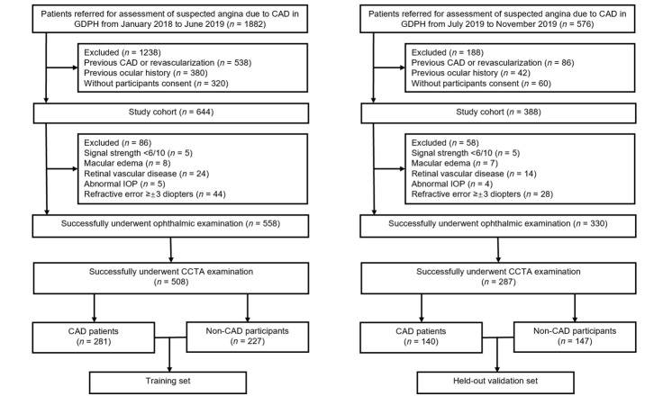
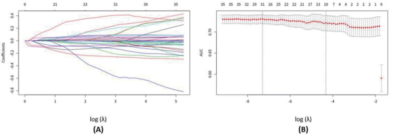
The prediction model consisting of 795 participants was developed in a training set of 508 participants with suspected angina due to CAD, and data were collected from January 2018 to June 2019. The held-out validation was conducted with 287 consecutive patients from July 2019 to November 2019. All patients with suspected CAD received optical coherence tomography angiography (OCTA) examination before undergoing coronary CT angiography. LASSO regression model was used for data reduction and feature selection. Multivariable logistic regression analysis was used to develop the retinal vasculature model for predicting the probability of the presence of CAD.

RESULTS

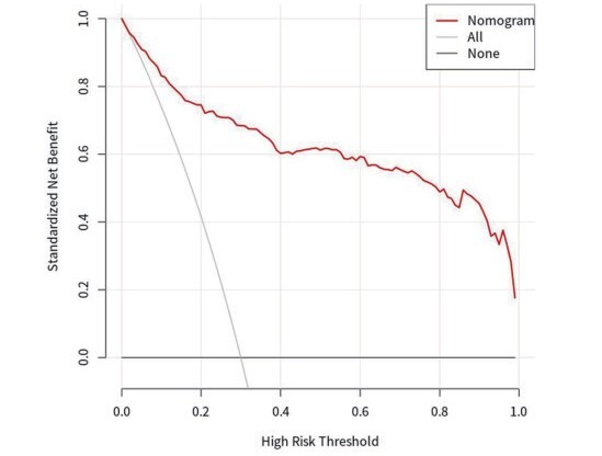
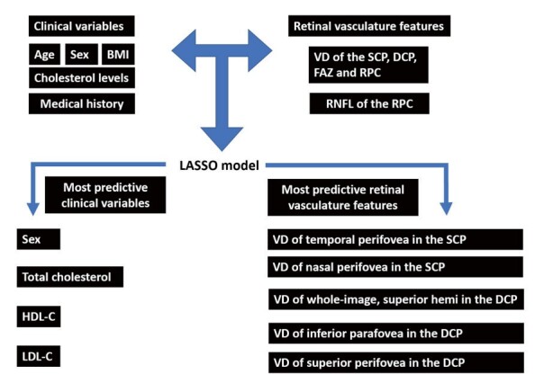
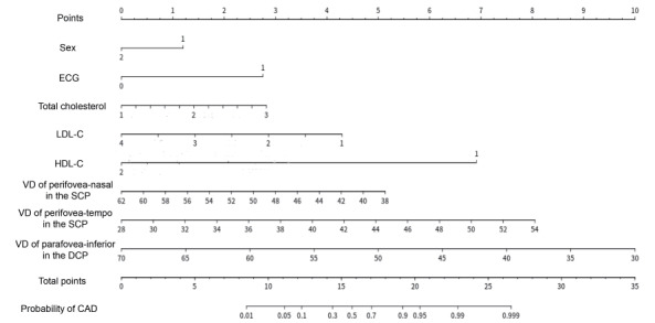
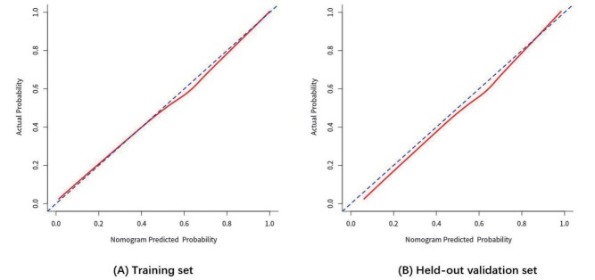
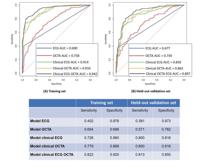
Three potential OCTA parameters including vessel density of the nasal and temporal perifovea in the superficial capillary plexus and vessel density of the inferior parafovea in the deep capillary plexus were further selected as independent retinal vasculature predictors. Model clinical electrocardiogram (ECG) OCTA (clinical variables+ECG+OCTA) was presented as the individual prediction nomogram, with good discrimination (AUC of 0.942 [95% CI, 0.923-0.961] and 0.897 [95% CI, 0.861-0.933] in the training and held-out validation sets, respectively) and good calibration. Decision curve analysis indicated the clinical applicability of this retinal vasculature nomogram.

CONCLUSIONS

The presented retinal vasculature nomogram based on individual probability can accurately identify the presence of CAD, which could improve patient selection and diagnostic yield of aggressive testing before determining a diagnosis.

摘要

目的

利用视网膜血管特征和临床变量开发并验证一个列线图,以预测疑似心绞痛患者的冠状动脉疾病(CAD)。

方法

该预测模型由 795 名参与者组成,其中 508 名参与者因 CAD 导致疑似心绞痛,数据收集于 2018 年 1 月至 2019 年 6 月。2019 年 7 月至 11 月,连续 287 名疑似 CAD 的患者进行了保留验证。所有疑似 CAD 的患者均在接受冠状动脉 CT 血管造影前接受了光学相干断层扫描血管造影(OCTA)检查。LASSO 回归模型用于数据缩减和特征选择。多变量逻辑回归分析用于开发预测 CAD 存在概率的视网膜血管模型。

结果

选择了三个潜在的 OCTA 参数,包括浅层毛细血管丛鼻侧和颞侧的血管密度以及深层毛细血管丛下旁中心凹的血管密度,作为独立的视网膜血管预测因子。模型临床心电图(ECG)OCTA(临床变量+ECG+OCTA)被呈现为个体预测列线图,具有良好的判别能力(在训练集和保留验证集中的 AUC 分别为 0.942[95%CI,0.923-0.961]和 0.897[95%CI,0.861-0.933])和良好的校准度。决策曲线分析表明了该视网膜血管列线图的临床适用性。

结论

该列线图基于个体概率,可以准确识别 CAD 的存在,这可以改善诊断前确定诊断时的患者选择和积极检测的诊断收益。

https://cdn.ncbi.nlm.nih.gov/pmc/blobs/2590/9135645/be69526317f9/29_62059_8.jpg
https://cdn.ncbi.nlm.nih.gov/pmc/blobs/2590/9135645/88155d3802f7/29_62059_1.jpg
https://cdn.ncbi.nlm.nih.gov/pmc/blobs/2590/9135645/9c9157657771/29_62059_2.jpg
https://cdn.ncbi.nlm.nih.gov/pmc/blobs/2590/9135645/393a06cc09c7/29_62059_3.jpg
https://cdn.ncbi.nlm.nih.gov/pmc/blobs/2590/9135645/3b3131f9db18/29_62059_4.jpg
https://cdn.ncbi.nlm.nih.gov/pmc/blobs/2590/9135645/0779559f1842/29_62059_5.jpg
https://cdn.ncbi.nlm.nih.gov/pmc/blobs/2590/9135645/0a1b511905e9/29_62059_6.jpg
https://cdn.ncbi.nlm.nih.gov/pmc/blobs/2590/9135645/f0da1364958a/29_62059_7.jpg
https://cdn.ncbi.nlm.nih.gov/pmc/blobs/2590/9135645/be69526317f9/29_62059_8.jpg

相似文献

1
Development and Validation of Retinal Vasculature Nomogram in Suspected Angina Due to Coronary Artery Disease.怀疑因冠状动脉疾病引起的心绞痛患者视网膜血管测图的制定与验证。
J Atheroscler Thromb. 2022 May 1;29(5):579-596. doi: 10.5551/jat.62059. Epub 2021 Mar 19.
2
Impaired retinal microcirculation in patients with non-obstructive coronary artery disease.非阻塞性冠状动脉疾病患者的视网膜微循环受损。
Microvasc Res. 2023 Jul;148:104533. doi: 10.1016/j.mvr.2023.104533. Epub 2023 Mar 31.
3
Retinal microvasculature impairments in patients with coronary artery disease: An optical coherence tomography angiography study.冠状动脉疾病患者的视网膜微血管损伤:一项光学相干断层扫描血管造影研究。
Acta Ophthalmol. 2022 Mar;100(2):225-233. doi: 10.1111/aos.14806. Epub 2021 Feb 24.
4
Development and validation of a risk prediction nomogram for in-stent restenosis in patients undergoing percutaneous coronary intervention.经皮冠状动脉介入治疗患者支架内再狭窄风险预测列线图的建立与验证。
BMC Cardiovasc Disord. 2021 Sep 14;21(1):435. doi: 10.1186/s12872-021-02255-4.
5
Retinal Microvasculature Changes in Patients With Coronary Total Occlusion on Optical Coherence Tomography Angiography.光学相干断层扫描血管造影术对冠状动脉完全闭塞患者视网膜微血管变化的研究
Front Med (Lausanne). 2021 Dec 16;8:708491. doi: 10.3389/fmed.2021.708491. eCollection 2021.
6
Quantitative assessment of retinal vessel density and thickness changes in internal carotid artery stenosis patients using optical coherence tomography angiography.采用光学相干断层血管造影术定量评估颈内动脉狭窄患者视网膜血管密度和厚度的变化。
Photodiagnosis Photodyn Ther. 2022 Sep;39:103006. doi: 10.1016/j.pdpdt.2022.103006. Epub 2022 Jul 12.
7
A model combining rest-only ECG-gated SPECT myocardial perfusion imaging and cardiovascular risk factors can effectively predict obstructive coronary artery disease.仅行静息状态心电图门控 SPECT 心肌灌注显像和心血管危险因素模型可有效预测阻塞性冠状动脉疾病。
BMC Cardiovasc Disord. 2022 Jun 15;22(1):268. doi: 10.1186/s12872-022-02712-8.
8
Early diabetic retinopathy diagnosis based on local retinal blood vessel analysis in optical coherence tomography angiography (OCTA) images.基于光学相干断层扫描血管造影(OCTA)图像中局部视网膜血管分析的早期糖尿病性视网膜病变诊断。
Med Phys. 2018 Oct;45(10):4582-4599. doi: 10.1002/mp.13142. Epub 2018 Sep 19.
9
Development and validation of a novel nomogram to predict chronic total occlusion before coronary angiography.一种用于在冠状动脉造影术前预测慢性完全闭塞的新型列线图的开发与验证
Eur Rev Med Pharmacol Sci. 2022 Nov;26(21):8011-8021. doi: 10.26355/eurrev_202211_30155.
10
A nomogram based on endothelial function and conventional risk factors predicts coronary artery disease in hypertensives.基于内皮功能和传统危险因素的列线图预测高血压患者的冠状动脉疾病。
BMC Cardiovasc Disord. 2023 Apr 28;23(1):217. doi: 10.1186/s12872-023-03235-6.

引用本文的文献

1
Retinal optical coherence tomography angiography (OCTA) biomarkers of cardiovascular disease: a review article.心血管疾病的视网膜光学相干断层扫描血管造影(OCTA)生物标志物:一篇综述文章。
Eye (Lond). 2025 May 14. doi: 10.1038/s41433-025-03780-8.
2
Retinal Microvascular Characteristics-Novel Risk Stratification in Cardiovascular Diseases.视网膜微血管特征——心血管疾病中的新型风险分层
Diagnostics (Basel). 2025 Apr 23;15(9):1073. doi: 10.3390/diagnostics15091073.
3
Coronary artery and retinal vascularization by optical coherence tomography angiography: are eyes the window to the heart?

本文引用的文献

1
Exercise Electrocardiography and Computed Tomography Coronary Angiography for Patients With Suspected Stable Angina Pectoris: A Post Hoc Analysis of the Randomized SCOT-HEART Trial.疑似稳定性心绞痛患者的运动心电图和计算机断层扫描冠状动脉造影:随机 SCOT-HEART 试验的事后分析。
JAMA Cardiol. 2020 Aug 1;5(8):920-928. doi: 10.1001/jamacardio.2020.1567.
2
Prediction of hypertension, hyperglycemia and dyslipidemia from retinal fundus photographs via deep learning: A cross-sectional study of chronic diseases in central China.基于深度学习的眼底照片对高血压、高血糖和血脂异常的预测:中国中部地区慢性病的横断面研究。
PLoS One. 2020 May 14;15(5):e0233166. doi: 10.1371/journal.pone.0233166. eCollection 2020.
3
光学相干断层扫描血管造影术对冠状动脉和视网膜血管成像:眼睛是心灵的窗口吗?
Graefes Arch Clin Exp Ophthalmol. 2025 Feb 15. doi: 10.1007/s00417-025-06769-x.
4
Artificial intelligence-based prediction of neurocardiovascular risk score from retinal swept-source optical coherence tomography-angiography.基于人工智能的视网膜扫频源光相干断层血管造影术预测神经心血管风险评分。
Sci Rep. 2024 Nov 7;14(1):27089. doi: 10.1038/s41598-024-78587-w.
5
Optical coherence tomography angiography in cardiovascular disease.光学相干断层扫描血管造影术在心血管疾病中的应用
Prog Cardiovasc Dis. 2024 Nov-Dec;87:60-72. doi: 10.1016/j.pcad.2024.10.011. Epub 2024 Oct 21.
6
Retinal Structural and Vascular Changes in Patients with Coronary Artery Disease: A Systematic Review and Meta-Analysis.冠状动脉疾病患者的视网膜结构和血管变化:系统评价与荟萃分析
Life (Basel). 2024 Mar 28;14(4):448. doi: 10.3390/life14040448.
7
Optical Coherence Tomography Angiography Changes in Patients Diagnosed With Acute Coronary Syndrome: A Systematic Review and Meta-Analysis.急性冠状动脉综合征患者的光学相干断层扫描血管造影变化:一项系统评价和荟萃分析
Cureus. 2024 Feb 13;16(2):e54121. doi: 10.7759/cureus.54121. eCollection 2024 Feb.
8
AI-integrated ocular imaging for predicting cardiovascular disease: advancements and future outlook.用于预测心血管疾病的人工智能集成眼部成像:进展与未来展望。
Eye (Lond). 2024 Feb;38(3):464-472. doi: 10.1038/s41433-023-02724-4. Epub 2023 Sep 14.
9
Development and validation of a nomogram for predicting significant coronary artery stenosis in suspected non-ST-segment elevation acute coronary artery syndrome with low-to-intermediate risk stratification.用于预测疑似非ST段抬高型急性冠状动脉综合征且风险分层为低至中度的严重冠状动脉狭窄的列线图的开发与验证
Front Cardiovasc Med. 2022 Dec 19;9:1013563. doi: 10.3389/fcvm.2022.1013563. eCollection 2022.
10
Retinal and Choroidal Thinning-A Predictor of Coronary Artery Occlusion?视网膜和脉络膜变薄——冠状动脉阻塞的一个预测指标?
Diagnostics (Basel). 2022 Aug 20;12(8):2016. doi: 10.3390/diagnostics12082016.
Association of Cardiovascular Mortality and Deep Learning-Funduscopic Atherosclerosis Score derived from Retinal Fundus Images.
心血管死亡率与基于眼底图像的深度学习眼底动脉硬化评分的相关性。
Am J Ophthalmol. 2020 Sep;217:121-130. doi: 10.1016/j.ajo.2020.03.027. Epub 2020 Mar 25.
4
Exploring the effect of hypertension on retinal microvasculature using deep learning on East Asian population.利用深度学习探究东亚人群高血压对视网膜微血管的影响。
PLoS One. 2020 Mar 5;15(3):e0230111. doi: 10.1371/journal.pone.0230111. eCollection 2020.
5
Machine learning of clinical variables and coronary artery calcium scoring for the prediction of obstructive coronary artery disease on coronary computed tomography angiography: analysis from the CONFIRM registry.基于临床变量和冠状动脉钙化评分的机器学习用于预测冠状动脉计算机断层扫描血管造影中的阻塞性冠状动脉疾病:来自CONFIRM注册研究的分析
Eur Heart J. 2020 Jan 14;41(3):359-367. doi: 10.1093/eurheartj/ehz565.
6
Retinal and choroidal vascular changes in coronary heart disease: an optical coherence tomography angiography study.冠心病患者视网膜和脉络膜血管变化:一项光学相干断层扫描血管造影研究
Biomed Opt Express. 2019 Mar 4;10(4):1532-1544. doi: 10.1364/BOE.10.001532. eCollection 2019 Apr 1.
7
Prediction of cardiovascular risk factors from retinal fundus photographs via deep learning.基于深度学习的眼底图像心血管风险因素预测。
Nat Biomed Eng. 2018 Mar;2(3):158-164. doi: 10.1038/s41551-018-0195-0. Epub 2018 Feb 19.
8
Unmasking Clever Hans predictors and assessing what machines really learn.揭开聪明汉斯预测者的面具,评估机器真正学到了什么。
Nat Commun. 2019 Mar 11;10(1):1096. doi: 10.1038/s41467-019-08987-4.
9
Coronary Microvascular Disease Pathogenic Mechanisms and Therapeutic Options: JACC State-of-the-Art Review.冠状动脉微血管疾病的发病机制和治疗选择:美国心脏病学会最新综述。
J Am Coll Cardiol. 2018 Nov 27;72(21):2625-2641. doi: 10.1016/j.jacc.2018.09.042.
10
The EYE-MI Pilot Study: A Prospective Acute Coronary Syndrome Cohort Evaluated With Retinal Optical Coherence Tomography Angiography.EYE-MI 初步研究:前瞻性急性冠脉综合征队列应用视网膜光学相干断层血管造影评估。
Invest Ophthalmol Vis Sci. 2018 Aug 1;59(10):4299-4306. doi: 10.1167/iovs.18-24090.