Department of Medicine, University of Fribourg, Chemin du Musée 8, 1700, Fribourg, CH, Switzerland.
Department of Radiology, Cantonal Hospital Fribourg, Fribourg, Switzerland.
Eur Radiol. 2021 Sep;31(9):6708-6716. doi: 10.1007/s00330-021-07842-9. Epub 2021 Mar 23.
To compare the impact of laxative enema preparation versus air/gas suction through a small catheter on image quality of prostate DWI.
In this single-center study, 200 consecutive patients (100 in each arm) with either enema or catheter preparation were retrospectively included. Two blinded readers independently assessed aspects of image quality on 5-point Likert scales. Scores were compared between groups and the influence of confounding factors evaluated using multivariable logistic regression. Prostate diameters were compared on DWI and T-weighted imaging using intraclass correlation coefficients.
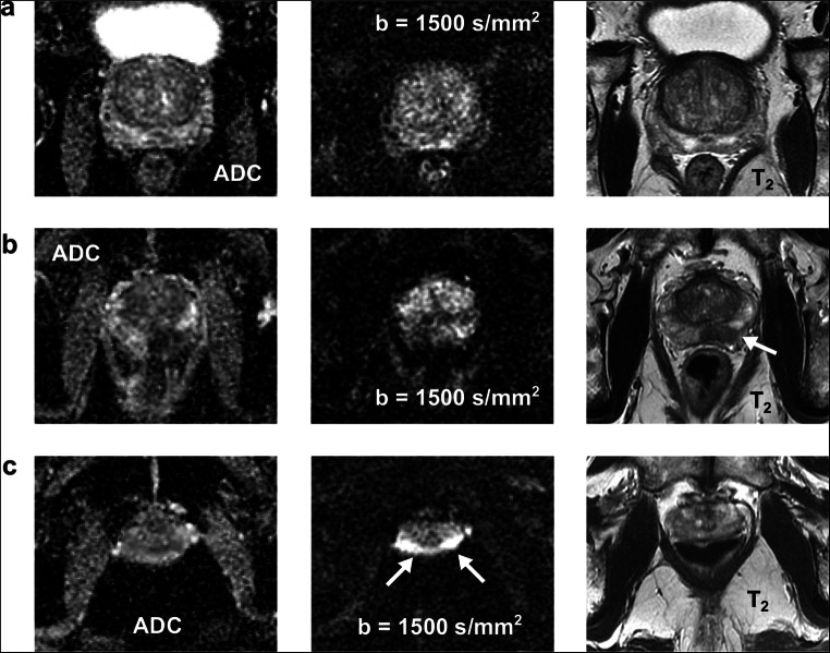
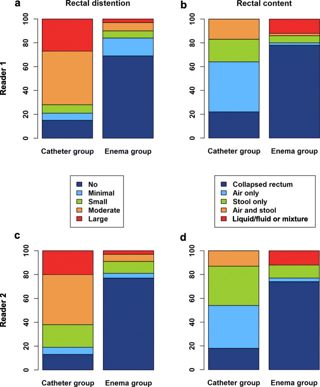
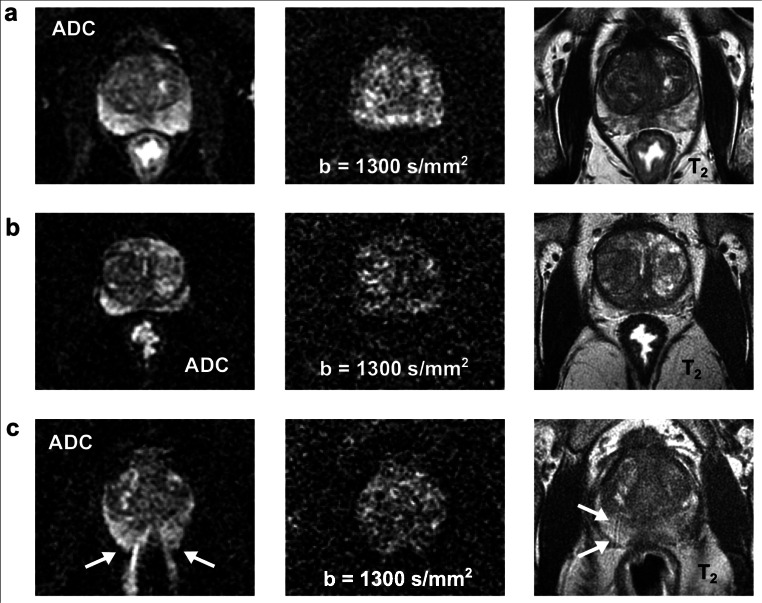
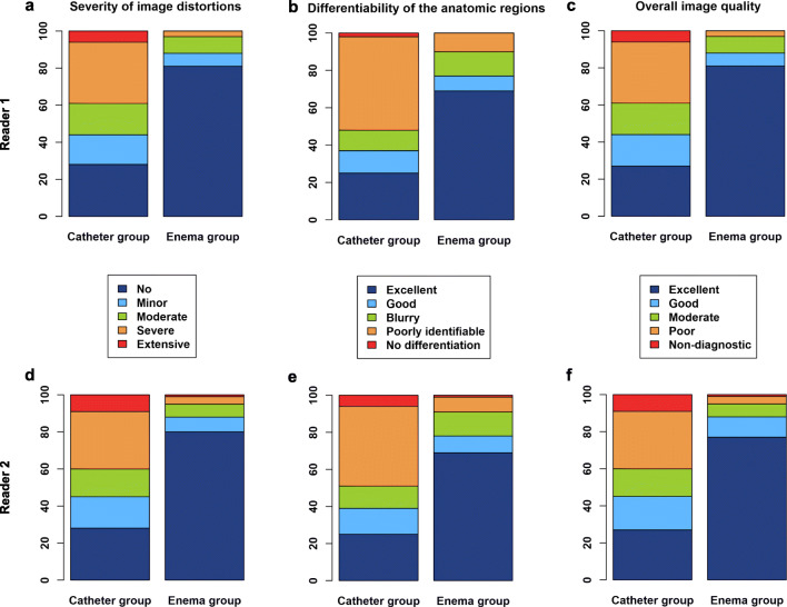
Image quality was significantly higher in the enema group regarding the severity of susceptibility-related artifacts (reader 1: 0.34 ± 0.77 vs. 1.73 ± 1.34, reader 2: 0.38 ± 0.86 vs. 1.76 ± 1.39), the differentiability of the anatomy (reader 1: 3.36 ± 1.05 vs. 2.08 ± 1.31, reader 2: 3.37 ± 1.05 vs. 2.09 ± 1.35), and the overall image quality (reader 1: 3.66 ± 0.77 vs. 2.26 ± 1.33, Reader 2: 3.59 ± 0.87 vs. 2.23 ± 1.38) with almost perfect inter-observer agreement (κ = 0.92-0.95). In the enema group, rectal distention was significantly lower and strongly correlated with the severity of artifacts (reader 1: ρ = 0.79, reader 2: ρ = 0.73). Furthermore, there were significantly fewer substantial image distortions, with odds ratios of 0.051 and 0.084 for the two readers which coincided with a higher agreement of the prostate diameters in the phase-encoding direction (0.96 vs. 0.89).
Enema preparation is superior to catheter preparation and yields substantial improvements in image quality.
• Enema preparation is superior to decompression of the rectum using air/gas suction through a small catheter. • Enema preparation markedly improves the image quality of prostate DWI regarding the severity of susceptibility-related artifacts, the differentiability of the anatomy, and the overall image quality and considerably reduces substantial artifacts that may impair a reliable diagnosis.
比较灌肠准备与小导管气/抽吸对前列腺 DWI 图像质量的影响。
本单中心研究回顾性纳入了 200 例(每组 100 例)接受灌肠或导管准备的连续患者。两名盲法读者独立使用 5 分 Likert 量表评估图像质量的各个方面。比较组间评分,并使用多变量逻辑回归评估混杂因素的影响。使用组内相关系数比较 DWI 和 T2 加权成像上的前列腺直径。
灌肠组在与磁化率相关的伪影严重程度(读者 1:0.34 ± 0.77 比 1.73 ± 1.34,读者 2:0.38 ± 0.86 比 1.76 ± 1.39)、解剖结构的可分辨性(读者 1:3.36 ± 1.05 比 2.08 ± 1.31,读者 2:3.37 ± 1.05 比 2.09 ± 1.35)和整体图像质量(读者 1:3.66 ± 0.77 比 2.26 ± 1.33,读者 2:3.59 ± 0.87 比 2.23 ± 1.38)方面显著更高,观察者间具有几乎完美的一致性(κ=0.92-0.95)。在灌肠组中,直肠扩张显著降低,与伪影严重程度呈强相关(读者 1:ρ=0.79,读者 2:ρ=0.73)。此外,明显较少出现实质性图像扭曲,两名读者的优势比分别为 0.051 和 0.084,这与相位编码方向上前列腺直径的一致性更高(0.96 比 0.89)一致。
灌肠准备优于通过小导管进行直肠减压,可显著改善图像质量。
• 灌肠准备优于通过小导管进行的空气/气体抽吸来排空直肠。• 灌肠准备显著改善了前列腺 DWI 的图像质量,降低了磁化率相关伪影的严重程度、解剖结构的可分辨性以及整体图像质量,大大减少了可能影响可靠诊断的实质性伪影。