Shao Jiahua, Zhu Jun, Chen Yi, Fu Qiwei, Li Lexiang, Ding Zheru, Wu Jun, Han Yaguang, Li Haobo, Qian Qirong, Zhou Yiqin
Department of Orthopedics, Shanghai Changzheng Hospital, Naval Medical University, Shanghai 200003, China.
Stem Cells Int. 2021 Mar 9;2021:6624874. doi: 10.1155/2021/6624874. eCollection 2021.
To evaluate the effect of Kartogenin-pretreated exosomes derived from infrapatellar fat pad mesenchymal stem cells on chondrocyte in vitro and articular cartilage regeneration in vivo.
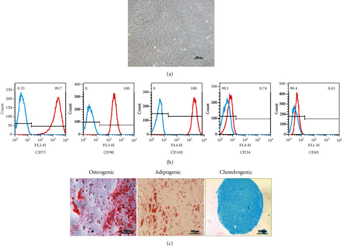
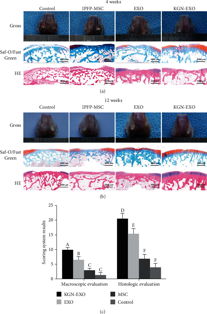
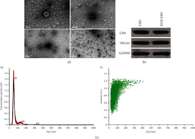
Infrapatellar fat pad mesenchymal stem cells (IPFP-MSCs) were isolated from rabbits to harvest exosomes. After identification of mesenchymal stem cells and exosomes, rabbit chondrocytes were divided into three groups for further treatment: the EXO group (chondrocytes treated with exosomes isolated from infrapatellar fat pad mesenchymal stem cells), KGN-EXO group (chondrocytes treated with exosomes isolated from infrapatellar fat pad mesenchymal stem cells pretreated with KGN), and control group. After processing and proliferation, phenotypic changes of chondrocytes were measured. In the in vivo study, 4 groups of rabbits with articular cartilage injury were treated with KGN-EXO, EXO, IPFP-MSCs, and control. Macroscopic evaluation and histological evaluation were made to figure out the different effects of the 4 groups on cartilage regeneration in vivo.
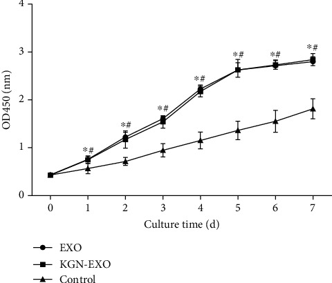
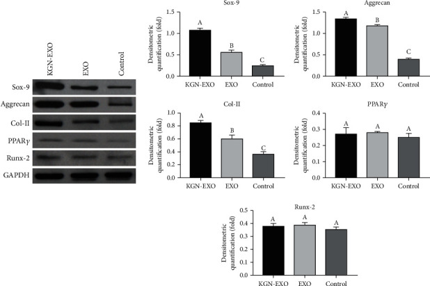
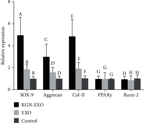
The proliferation rate of chondrocytes in the EXO or KGN-EXO group was significantly higher than that in the control group ( < 0.05). The qRT-PCR results showed that the expression of Sox-9, Aggrecan, and Col II was the highest in the KGN-EXO group compared with the EXO group and the control group ( < 0.05). The results of Western blot were consistent with the results of qRT-PCR. In vivo, the cartilage defects in the KGN-EXO group showed better gross appearance and improved histological score than those in IPFP-MSC groups, EXO groups, and control groups ( < 0.05). At 12 weeks, the defect site in the KGN-EXO group was almost completely repaired with a flat and smooth surface, while a large amount of hyaline cartilage-like structures and no obvious cracks were observed.
Our study demonstrates that the exosomes isolated from infrapatellar fat pad mesenchymal stem cells pretreated with KGN have potent ability to induce chondrogenic differentiation of stem cells, effectively promoting the proliferation and the expression of chondrogenic proteins and genes of chondrocytes. The KGN-EXO can also promote the repair of articular cartilage defects more effectively, which can be used as a potential therapeutic method in the future.
评估经Kartogenin预处理的髌下脂肪垫间充质干细胞来源的外泌体对体外软骨细胞及体内关节软骨再生的影响。
从兔体内分离髌下脂肪垫间充质干细胞(IPFP-MSCs)以获取外泌体。在鉴定间充质干细胞和外泌体后,将兔软骨细胞分为三组进行进一步处理:EXO组(用从髌下脂肪垫间充质干细胞分离的外泌体处理的软骨细胞)、KGN-EXO组(用经KGN预处理的髌下脂肪垫间充质干细胞分离的外泌体处理的软骨细胞)和对照组。经过处理和增殖后,检测软骨细胞的表型变化。在体内研究中,对4组关节软骨损伤的兔分别用KGN-EXO、EXO、IPFP-MSCs和对照进行处理。进行宏观评估和组织学评估以明确4组对体内软骨再生的不同影响。
EXO组或KGN-EXO组软骨细胞的增殖率显著高于对照组(<0.05)。qRT-PCR结果显示,与EXO组和对照组相比,KGN-EXO组中Sox-9、聚集蛋白聚糖和Ⅱ型胶原的表达最高(<0.05)。蛋白质免疫印迹结果与qRT-PCR结果一致。在体内,KGN-EXO组的软骨缺损在大体外观上显示出比IPFP-MSC组、EXO组和对照组更好的表现,组织学评分也有所改善(<0.05)。在12周时,KGN-EXO组的缺损部位几乎完全修复,表面平坦光滑,观察到大量透明软骨样结构且无明显裂缝。
我们的研究表明,经KGN预处理的髌下脂肪垫间充质干细胞分离的外泌体具有诱导干细胞软骨分化的强大能力,能有效促进软骨细胞的增殖以及软骨生成蛋白和基因的表达。KGN-EXO还能更有效地促进关节软骨缺损的修复,有望在未来用作一种潜在的治疗方法。