Huang Jingyan, Li Ruoqi, Yang Jinghong, Cai Min, Lee Yichen, Wang Anxun, Cheng Bin, Wang Yan
Hospital of Stomatology, Guanghua School of Stomatology, Sun Yat-sen University and Guangdong Key Laboratory of Stomatology, Guangzhou, Guangdong, 510055, China.
The First Affiliated Hospital of Sun Yat-sen University, Guangzhou, Guangdong, 510080, China.
Bioact Mater. 2021 Mar 10;6(10):3164-3176. doi: 10.1016/j.bioactmat.2021.02.023. eCollection 2021 Oct.
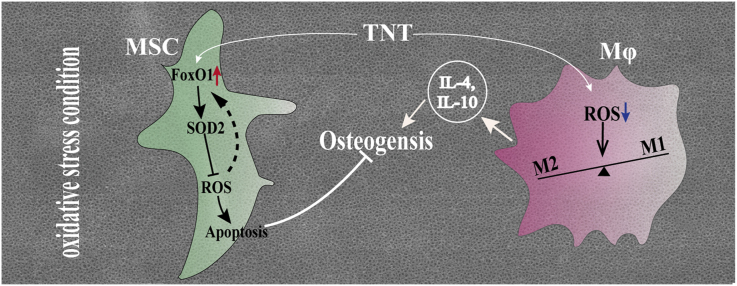
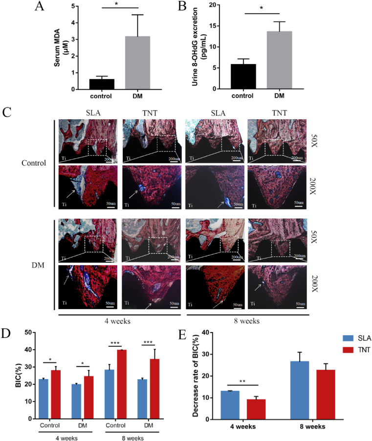
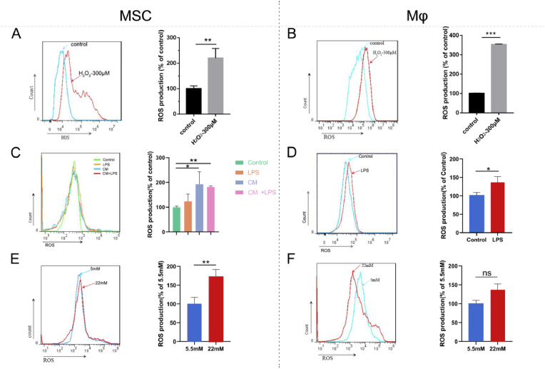
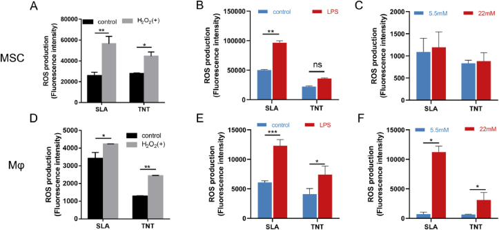
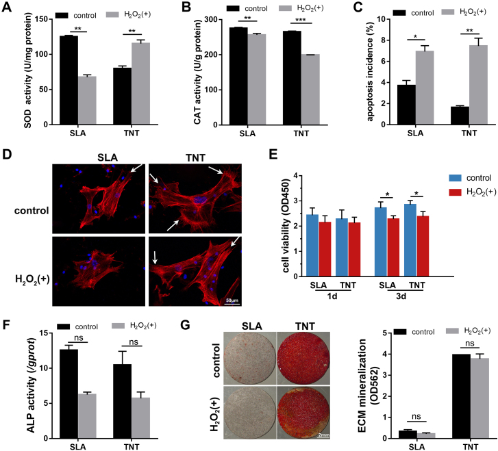
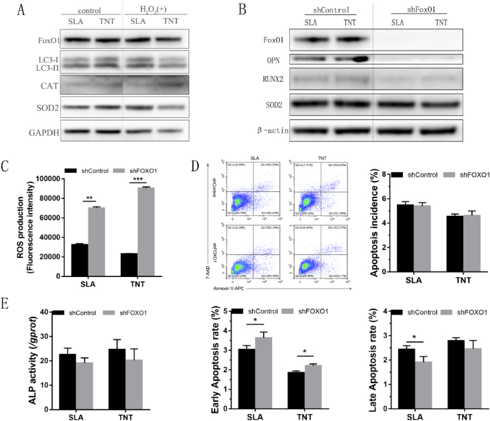
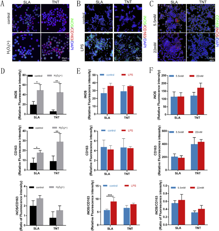
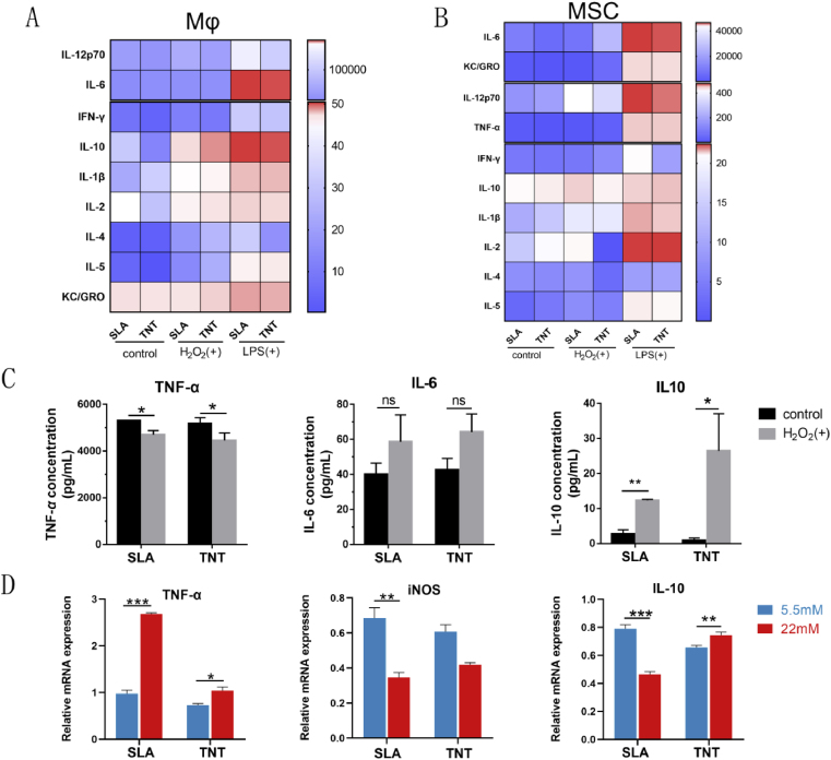
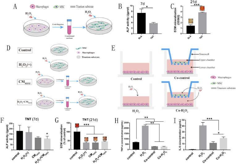
Varieties of pathological conditions, including diabetes, are closely related to oxidative stress (OS), but the osseointegration or bioadaptation of implants to OS and the related mechanism remain poorly explored. In this study, the antioxidation and osteoimmune regeneration of titanium implants with micro/nanotopographies were evaluated under HO-, lipopolysaccharide (LPS)- and hyperglycemia-mediated cellular OS models and in diabetic rats as a representative animal model of OS. TiO nanotube (TNT) coating on titanium implants directly induced superior osteogenic differentiation of bone mesenchymal stem cells (MSCs) and osseointegration compared with microscale sand blasted-acid etched topography (SLA) under OS, attributed to higher superoxide dismutase 2 activity, the neutralization of intracellular reactive oxygen species (ROS), and less apoptosis. Mechanistically, the oxidation resistance on TNT is driven by upregulated forkhead box transcription factor O1 (FoxO1), which is abolished after knockdown of FoxO1 via shRNA in MSCs. Indirectly, TNT also alleviates OS in macrophages, therefore inducing a higher portion of the M2 phenotype under OS with increased secretion of the anti-inflammatory cytokine IL-10, further promoting the osseoimmunity capacity compared with SLA. The current study not only suggests the potential application of TiO nanotube-coated titanium implants in compromised conditions but also provides a systematic evaluation strategy for the future development of bone biomaterials.
包括糖尿病在内的多种病理状况都与氧化应激(OS)密切相关,但植入物对氧化应激的骨整合或生物适应性及其相关机制仍未得到充分探索。在本研究中,我们在过氧化氢(HO-)、脂多糖(LPS)和高血糖介导的细胞氧化应激模型以及作为氧化应激代表性动物模型的糖尿病大鼠中,评估了具有微/纳米拓扑结构的钛植入物的抗氧化和骨免疫再生能力。与微尺度喷砂-酸蚀形貌(SLA)相比,钛植入物上的TiO纳米管(TNT)涂层在氧化应激条件下直接诱导了骨间充质干细胞(MSCs)更好的成骨分化和骨整合,这归因于更高的超氧化物歧化酶2活性、细胞内活性氧(ROS)的中和以及更少的细胞凋亡。从机制上讲,TNT的抗氧化能力是由上调的叉头框转录因子O1(FoxO1)驱动的,在通过shRNA敲低MSCs中的FoxO1后,这种能力被消除。间接而言,TNT还减轻了巨噬细胞中的氧化应激,因此在氧化应激条件下诱导出更高比例的M2表型,抗炎细胞因子IL-10的分泌增加,与SLA相比进一步提高了骨免疫能力。本研究不仅表明了TiO纳米管涂层钛植入物在受损条件下的潜在应用,还为骨生物材料的未来发展提供了一种系统的评估策略。