Rawji Vishal, Kaczmarczyk Isabella, Rocchi Lorenzo, Fong Po-Yu, Rothwell John C, Sharma Nikhil
Department of Clinical and Movement Neurosciences, Queen Square Institute of Neurology, University College London, London WC1N 3BG, UK.
Department of Medical Sciences and Public Health, University of Cagliari, 09124 Cagliari, Italy.
Brain Sci. 2021 Mar 4;11(3):326. doi: 10.3390/brainsci11030326.
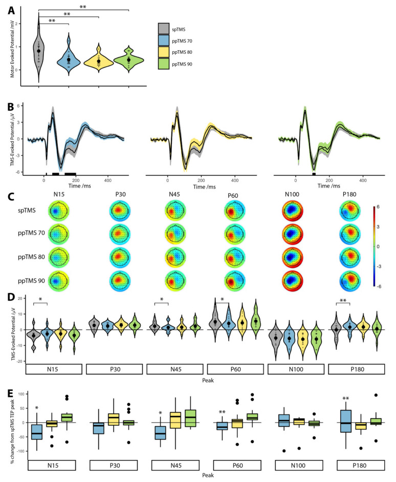
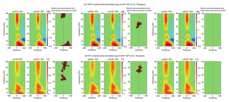
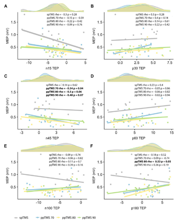
Motor cortex (M1) paired-pulse TMS (ppTMS) probes excitatory and inhibitory intracortical dynamics by measurement of motor-evoked potentials (MEPs). However, MEPs reflect cortical and spinal excitabilities and therefore cannot isolate cortical function. Concurrent TMS-EEG has the ability to measure cortical function, while limiting peripheral confounds; TMS stimulates M1, whilst EEG acts as the readout: the TMS-evoked potential (TEP). Whilst varying preconditioning stimulus intensity influences intracortical inhibition measured by MEPs, the effects on TEPs is undefined. TMS was delivered to the left M1 using single-pulse and three, ppTMS paradigms, each using a different preconditioning stimulus: 70%, 80% or 90% of resting motor threshold. Corticospinal inhibition was present in all three ppTMS conditions. ppTMS TEP peaks were reduced predominantly under the ppTMS 70 protocol but less so for ppTMS 80 and not at all for ppTMS 90. There was a significant negative correlation between MEPs and N45 TEP peak for ppTMS 70 reaching statistical trends for ppTMS 80 and 90. Whilst ppTMS MEPs show inhibition across a range of preconditioning stimulus intensities, ppTMS TEPs do not. TEPs after M1 ppTMS vary as a function of preconditioning stimulus intensity: smaller preconditioning stimulus intensities result in better discriminability between conditioned and unconditioned TEPs. We recommend that preconditioning stimulus intensity should be minimized when using ppTMS to probe intracortical inhibition.
运动皮层(M1)配对脉冲经颅磁刺激(ppTMS)通过测量运动诱发电位(MEP)来探究兴奋性和抑制性皮质内动力学。然而,MEP反映的是皮质和脊髓的兴奋性,因此无法分离皮质功能。同步经颅磁刺激-脑电图(TMS-EEG)有能力测量皮质功能,同时限制外周干扰;TMS刺激M1,而EEG作为读出信号:即TMS诱发电位(TEP)。虽然改变预处理刺激强度会影响通过MEP测量的皮质内抑制,但对TEP的影响尚不清楚。使用单脉冲和三种ppTMS范式将TMS施加到左侧M1,每种范式使用不同的预处理刺激:静息运动阈值的70%、80%或90%。在所有三种ppTMS条件下均存在皮质脊髓抑制。ppTMS的TEP峰值在ppTMS 70方案下主要降低,但在ppTMS 80时降低较少,在ppTMS 90时则完全没有降低。对于ppTMS 70,MEP与N45 TEP峰值之间存在显著负相关,对于ppTMS 80和90达到统计趋势。虽然ppTMS的MEP在一系列预处理刺激强度下均显示出抑制作用,但ppTMS的TEP并非如此。M1 ppTMS后的TEP随预处理刺激强度而变化:较小的预处理刺激强度导致条件性和非条件性TEP之间的可辨别性更好。我们建议在使用ppTMS探究皮质内抑制时,应将预处理刺激强度降至最低。